

The first Testosterone Kabbalah reached almost 10 million views. I kept studying the subject after I published it. This is the version I wanted to be able to write then.
They control everything. They stole your testosterone. Your testosterone was promised to them 3,000 years ago.
The joke hides a real problem. Men are taught to treat testosterone as a number that can be pushed upward. The blood value is only the visible output of a timed biological system. Circadian rhythm, sleep, energy availability, thyroid state, reproductive signaling, Leydig-cell function, mitochondrial cholesterol transport, steroid enzymes, liver handling, local conversion, and receptor response all shape the result.
A low result tells you what was present in that blood sample. It does not tell you why. Luteinizing hormone, or LH, follicle-stimulating hormone, or FSH, sex hormone-binding globulin, or SHBG, free testosterone, prolactin, sleep timing, health, and exposure history help locate the cause. A normal total-testosterone result can still coexist with impaired fertility, weak tissue response, sexual dysfunction, or poor recovery.
Everything depends on identifying the part of the system that limits the result. Once that is clear, the order of action becomes much easier.
Contents
Opening. Testosterone boosters, diagnosis, and the systems map
Before the map: what testosterone boosters actually change
Human evidence, animal evidence, product identity, adulteration, and the difference between symptom relief, temporary output, and restoration
Fadogia, Bulbine, Tongkat ali, ashwagandha, shilajit, fenugreek, zinc, boron, Tribulus, Butea superba, akarkara, D-aspartic acid, and complex formulas
Products that target LH, estrogen, DHT, free testosterone, libido, erections, or training performance
Start here: find the limiting layer
Define the problem, record the changed context, screen urgent findings, and decide whether to stabilize, test, or escalate
The Testosterone Kabbalah systems map
Timing, sleep, nutrition, thyroid function, mitochondria, cholesterol transport, reproductive signaling, steroid enzymes, liver and gut handling, tissue conversion, and receptor action
Part I. How the system works
1. A testosterone result has several layers
Production, blood concentration, binding, local conversion, receptor response, symptoms, fertility, and durable function
2. Testosterone rises through sleep cycles
Sleep onset, slow-wave sleep, REM, continuity, circadian timing, testing time, and the distinct clocks of pituitary hormones
Sleep opportunity, airway resistance, apnea, nasal obstruction, allergies, mouth taping, nasal strips, nasal rinsing, and sleep tracking
3. Retinal light, dopamine, and blue light
Morning and daytime outdoor light, evening blue-rich light, retinal dopamine, circadian contrast, bromantane, and seasonal context
4. Energy availability
Calorie intake, glycogen, fasting, dieting, illness, training load, and low energy availability
5. Nutrition, body composition, and the supplement reference
Evidence hierarchy, the original Ray Peat sample food pyramid, energy context, carbohydrate, protein, gelatin, dietary fat, cholesterol, and PUFA
Salt, thiamine, iodine, selenium, magnesium, zinc, vitamins A, D, E and K, P5P, glycine, taurine, TMG, inositol, and other layer-specific tools
Supplements arranged by circadian rhythm and sleep, thyroid function, mitochondrial energy, prolactin and estrogen, gut function, liver clearance, body composition, fertility, and testicular function
Orange juice flavanones, raw carrot salad, white button mushrooms, bamboo shoots, intestinal tolerance, aromatase, estrogen handling, insulin resistance, adiposity, and SHBG
6. Training and recovery
Resistance training, aerobic work, intervals, endurance, adaptation, recovery signals, and deloading
7. Thyroid and metabolic state
Thyroid output, mitochondrial respiration, metabolic capacity, binding proteins, transport, and peripheral T4-to-T3 conversion
8. Steroid production
Brain-to-testis signaling, Ray Peat’s hierarchy, LH and Leydig cells, FSH and Sertoli cells, cholesterol supply, StAR transport, and mitochondrial conversion
Pregnenolone, the delta-5 and delta-4 routes, testosterone synthesis, and intratesticular testosterone
9. Liver, gut, serotonin, prolactin, illness, and stress
SHBG, hormone metabolism and clearance, bile, endotoxin, gut serotonin, motility, inflammation, glucocorticoids, and active illness
Prolactin, dopamine, pornography, compulsive masturbation, sexual context, and the difference between repeated novelty and partnered sex
10. Conversion, transport, and tissue response
Pregnenolone, progesterone, DHT, 5-alpha-reductase, estradiol, aromatase, SHBG, free testosterone, androgen receptors, symptoms, and tissue function
Part II. Read the laboratory pattern
11. Laboratory interpretation
A valid baseline, matched collection conditions, the core laboratory set, and the difference between serum testosterone, intratesticular testosterone, metabolites, and tissue action
LH beside testosterone, the LH-to-testosterone relationship, INSL3, Leydig-cell capacity, FSH, prolactin, estradiol, SHBG, thyroid markers, and fertility testing
Seven endocrine patterns, from central suppression and primary testicular failure to binding-protein discordance, exogenous exposure, and persistent symptoms with valid androgen values
12. Causes, exposures, and age
Testicular, pituitary, hypothalamic, medication, substance, environmental, occupational, and anabolic-androgenic steroid causes
Aging as accumulated metabolic, sleep, exposure, illness, and behavioral burden rather than an isolated mechanism
Part III. Restore the system
13. The natural restoration plan
Gate 1. Validate and classify: Complete the intake, standardize measurement, confirm persistence, and locate the limiting layer
Gate 2. Stabilize circadian rhythm and sleep: Fix wake timing, build day-night light contrast, protect complete sleep cycles, and screen airway and sleep disruptors
Gate 3. Restore energetic and metabolic conditions: Reconstruct the energy history, correct under-fueling, reduce excess adiposity without crash dieting, and correct defined nutrient needs
Gate 4. Adjust training and remove suppressors: Match training to recovery, review drugs and hormones, stop compulsive habits, and treat active illness or gut disease
Gate 5. Retest, decide, and preserve options: Hold the intervention long enough, repeat matched testing, escalate unresolved patterns, and define the medical branch when needed
14. Daily and weekly practice
Light, meals, training, sleep, airway care, supplements, symptom tracking, and the weekly review
15. Monitor the result
Process measures, outcome measures, reassessment intervals, comparable testing conditions, several levels of success, and predefined failure criteria
16. Pattern-based troubleshooting
Poor sleep, obesity and low SHBG, energy deficit, high LH, low LH with high prolactin, persistent symptoms with normal testosterone, suppressed LH with high testosterone, and numerical improvement without functional recovery
17. Where the natural approach ends
Persistent central, testicular, fertility, structural, medication-related, or systemic disease patterns that require direct medical assessment
Final decision and reference material
The final decision rule: Locate the first weak layer, change the smallest useful variable, and judge durable function under matched conditions
References: Studies, patents, official Ray Peat articles, and official Georgi Dinkov articles
Before the map: what testosterone boosters actually change
A product sold as a testosterone booster may act on testosterone, another hormone, a binding protein, blood flow, arousal, stress, sleep, training performance, or nothing measurable. Those outcomes are often sold under the same name.
Five different effects need separate labels.
Deficiency correction replaces something the body lacks. Zinc can help when zinc deficiency is limiting reproductive function. More zinc after sufficiency does not keep raising testosterone.[1]
Signal stimulation pushes an instruction sent to the testes. A substance that raises luteinizing hormone, usually shortened to LH, increases testicular demand while it is present. The effect depends on the Leydig cells being able to answer.
Measurement change alters the relationship between total and free testosterone. Sex hormone-binding globulin, or SHBG, carries testosterone in blood. Lower SHBG can make calculated free testosterone look higher without increasing production.
Symptom change improves libido, erections, mood, or training through another pathway. Better blood flow can improve an erection while testosterone remains unchanged.
Temporary output raises a value during use. Durable restoration means that the limiting cause has improved and the system still works after the push is removed. Most booster trials never test that question.
A booster can push the output while leaving the machinery unchanged.
Human evidence, animal evidence, and product evidence
Product identity matters. The label on the bottle is only the beginning. Plant species, plant part, extraction method, standardized constituent, dose, and product authenticity all matter.
Tongkat ali, longjack, and Malaysian ginseng refer to Eurycoma longifolia. Physta and LJ100 are named root extracts. Evidence from either extract applies to that preparation, not every tongkat powder.
The same rule applies across the list. Akarkara, akalkara, and pellitory refer to Anacyclus pyrethrum. Ashwagandha is Withania somnifera. Fenugreek is Trigonella foenum-graecum. Testofen and Furosap are named fenugreek seed extracts. Maca is Lepidium meyenii. Shilajit is a variable mineral-organic material, and PrimaVie is a purified branded ingredient. These distinctions decide whether a study tested the product in the reader’s hand.
The human record is small. A 2021 review found 32 randomized herbal trials covering 13 plants. Nine trials reported a significant testosterone increase, and only six were judged at low risk of bias.[2] A later review included 52 studies. Most products did not raise total testosterone. Its possibly effective label required only one positive study. The studies behind that category were small, industry-funded, and at high risk of bias. Only eight of the 52 studies reported adverse effects. These trials measured output during treatment. They left durable recovery after withdrawal unanswered.[3]
Products with no convincing human restoration evidence
Fadogia agrestis became popular from a short rat experiment that raised testosterone and sexual behavior. A later study gave rats an aqueous stem extract at the same doses for 28 days. It changed several testicular measures at every dose. Testicular glutamate dehydrogenase, used in that paper as a marker of mitochondrial injury, fell as the dose rose. Ten days after withdrawal, several measures were still abnormal, especially at 50 and 100 mg/kg.[4][5] The published evidence remains animal-only, with a testicular safety signal and no controlled human efficacy or long-term safety trial.
Bulbine natalensis also rests on rat studies of an aqueous stem extract. After seven days, 25 and 50 mg/kg raised testosterone, LH, FSH, and mating success. At 100 mg/kg, testosterone fell and mating success was worse. A separate 14-day study found altered liver and kidney markers and mild tissue damage.[6][7] The dose reversal and organ findings prevent any safe human dose from being derived from these experiments.
Akarkara improved sexual behavior and accessory-organ measures in rats given a petroleum-ether root extract.[8] The evidence applies to that laboratory extract in rats. Ordinary root powder has no controlled human testosterone trial behind it.
Tribulus terrestris has been tested in men. Controlled trials found no reliable increase in testosterone, androstenedione, or LH.[9][10] A 2025 review found no consistent androgen effect across ten studies.[11] Some sexual-function studies reported improvement. An erectile or libido result remains a different endpoint from testosterone production.
Butea superba has a small erectile-dysfunction trial, one case report of very high dihydrotestosterone, and a later erectile trial whose first active batch was suspected of containing a phosphodiesterase-5 inhibitor.[12][13][14] Dihydrotestosterone is shortened to DHT. It is a potent androgen made from testosterone in many tissues. This record supports neither dependable endocrine restoration nor routine use.
I found no useful effect from Butea superba, Tongkat ali, Fadogia agrestis, Bulbine natalensis, or Akarkara in my own use. That experience led me to audit these products more carefully. Human trials and toxicology determine the published conclusion.
Products with limited or extract-specific human signals
Tongkat ali has the strongest human testosterone signal among the frequently marketed herbs. A 2022 meta-analysis found a higher average total testosterone result, with the clearest signal in men classified as hypogonadal.[15] Most studies used a named standardized extract. Samples were small, follow-up was short, and free testosterone, LH, follicle-stimulating hormone, symptoms, and persistence after stopping were reported less consistently.
One twelve-week Physta trial in older men increased total testosterone. The free-testosterone result was less clear, and the manufacturer funded the study and employed two authors.[16] A short trial in young men increased total and free testosterone and estradiol without changing LH or follicle-stimulating hormone.[17] Tongkat ali is conditional, extract-specific support. It is the wrong tool for sleep apnea, pituitary disease, primary testicular failure, or continued androgen suppression.
Ashwagandha uses several non-equivalent extracts, including KSM-66, Sensoril, and Shoden. One small crossover trial in overweight men found higher salivary testosterone and dehydroepiandrosterone sulfate, usually shortened to DHEA-S. Fatigue, vigor, sexual well-being, and estradiol did not improve beyond placebo.[18] The result applies to that extract, group, and endpoint.
Fenugreek trials use Testofen, Furosap, and other standardized seed extracts. A 2024 randomized trial found no significant pooled advantage over placebo for plasma total testosterone or the free-testosterone index. Salivary testosterone changed, and one dose comparison affected the index.[19] Culinary fenugreek is a different preparation and should not inherit the extract claim.
Purified shilajit raised total and free testosterone in one ninety-day trial of healthy men aged 45 to 55.[20] The claim belongs to that purified ingredient and that population. Independent replication, contaminant control, long-term safety, and persistence remain limited. Raw or unpurified material is a separate product.
These findings support product-specific conclusions. They give no reason for a default stack.
Nutrients only help when the nutrient is limiting
Correct a real deficiency. Zinc, magnesium, vitamin D, iodine, selenium, vitamins A and E, and B vitamins support normal physiology. Deficiency can impair health and reproductive function. Replacement can remove that constraint.
Sufficiency changes the rule. High-dose zinc can cause copper deficiency. Too much iodine or selenium can damage thyroid function. Fat-soluble vitamins can accumulate. Magnesium may help sleep or correct low intake without directly restoring testosterone. Randomized vitamin D trials have not shown a reliable testosterone increase in men who started with normal testosterone.[21]
Small studies do not create a protocol. Boron has changed SHBG or steroid measurements in small, short studies. That is a measurement signal, not evidence of durable production or clinical benefit. ZMA combines zinc, magnesium, and vitamin B6. It inherits the same deficiency logic.
A nutrient enters the plan when diet, symptoms, disease, or measurement establishes a plausible need. A broad micronutrient stack makes it harder to learn which input mattered.
A one-month high-output experiment
In 2013, Georgi Dinkov described a self-experiment built after two years of reading and testing. The first account named 140 grams of protein per day. The daily total was split into two servings of 20 grams of whey, 20 grams of casein, and 30 grams of gelatin, followed by orange juice. The supplement list gave 30 milligrams of zinc, 5 milligrams of vitamin B6, 2,000 milligrams of mixed tocopherols, 3,500 milligrams of branched-chain amino acids, and magnesium without a stated dose.
Georgi Dinkov reported total testosterone above the laboratory’s upper measurement limit of 1,500 ng/dL. No laboratory report, baseline panel, control period, or independent replication accompanied the post. The result remains a self-experiment.
In 2014, Georgi Dinkov described the month in more detail. He said his usual diet was high in protein and sugar and that many of his foods supplied substantial cholesterol. The expanded account added 15 grams of taurine, 6 to 7 grams of aspirin, 2,500 milligrams of mixed tocopherols, 45 milligrams of vitamin K2 as MK-4, 30 milligrams of zinc, 10 milligrams of vitamin B6, and 1,500 milligrams of thiamine. It also included branched-chain amino acids at a dose he did not remember, 1,500 milligrams of tyrosine with each branched-chain amino-acid dose, 200 to 400 milligrams of caffeine, and niacinamide that sometimes reached 8 grams per day.
The two accounts differ. The later account gives 2,500 rather than 2,000 milligrams of vitamin E, 10 rather than 5 milligrams of vitamin B6, and no exact branched-chain amino-acid dose. Those differences matter. They cannot be silently combined into one precise recipe.
The logic of the experiment is more useful than the final number:
Substrate and energy. Sugar supplied carbohydrate. Thiamine and niacinamide were used to push glucose oxidation and cellular energy. Caffeine raised metabolic drive. Cholesterol supplied one source of steroid substrate.
Amino-acid balance. Gelatin lowered the relative tryptophan and cysteine load of the protein mixture. Branched-chain amino acids compete with tryptophan for transport into the brain. They also compete with tyrosine, which explains why tyrosine was added. Vitamin B6 supports aromatic amino-acid decarboxylation. The intended direction was less serotonin, more dopamine, and less prolactin.
Stress and steroidogenic conditions. Aspirin, vitamin E, taurine, vitamin K2, magnesium, and zinc were used to change prostaglandin signaling, lipid oxidation, inhibitory signaling, mineral status, and the environment in which steroidogenesis occurs.
Every layer changed at once. The result therefore belongs to the combined state. It cannot identify which ingredient mattered, whether the value would persist after stopping, or whether a man with a different limiting layer would respond.
Georgi Dinkov later called the expanded schedule “not a healthy schedule” and said it was done purely as an experiment. That warning is justified. Aspirin at 6 to 7 grams per day can cause salicylate toxicity, bleeding, acid-base disturbance, and kidney injury. Niacinamide at several grams per day can cause dose-dependent adverse effects, including gastrointestinal and liver injury. Very high vitamin E can further inhibit platelet function when combined with aspirin.[22][23][24] Caffeine can also damage the sleep foundation this article places first.[25]
This experiment shows how several metabolic and signaling layers can be pushed together. The natural protocol below excludes it.
Products that target LH, estrogen, DHT, or free testosterone
LH. D-aspartic acid is marketed as an LH signal. In a twelve-week randomized trial of resistance-trained men, it did not improve total testosterone, free testosterone, estradiol, body composition, or training outcomes.[26]
Estradiol. Aromatase-oriented products try to reduce conversion of testosterone into estradiol. Estradiol is an estrogen that men also need for bone, body composition, sexual function, and hormonal feedback.[27] Lowering it can raise LH and testosterone during use by weakening feedback. That is hormone manipulation. It can create a new deficiency while the original cause remains.
DHT, prolactin, and receptors. Products sold as DHT boosters, 5-alpha-reductase boosters, prolactin reducers, or androgen-receptor sensitizers must be judged by the human outcome they produce. An enzyme change matters only when it improves the relevant function without creating a new hormonal problem.
Prescription stimulation. Drugs that raise LH, activate the LH receptor, lower prolactin, or inhibit aromatase can be useful for defined medical patterns. They need a diagnosis, monitoring, and a fertility plan. They are not natural restoration tools.
Libido, erections, performance, and laboratory spikes
Sexual function. Maca improved symptom scores in one randomized trial without a significant advantage for total or free testosterone.[28] That can still be a useful sexual outcome. It remains separate from endocrine restoration.
Blood flow and performance. Arginine, citrulline, yohimbine, horny goat weed, and products containing phosphodiesterase-5-like compounds mainly target blood flow or arousal. Creatine has established performance uses. In one crossover study of 20 young rugby players, testosterone did not change. DHT rose from 0.98 to 1.53 nmol/L after seven days of loading and remained at 1.38 nmol/L after 14 days of maintenance.[29] Treat that as one short serum finding. A reproducible DHT or tissue-effect protocol requires replication.
Training. Hard resistance exercise, sprints, and intervals can cause short hormone changes. Training improves strength, fitness, insulin sensitivity, and body composition. The brief post-workout rise does not explain chronic muscle growth or restored baseline testosterone.[30][31]
Cold and sun exposure. Cold showers, ice baths, testicular icing, and genital sun exposure lack controlled human evidence for durable restoration. Daylight reaching the eyes has a clear role in setting circadian rhythm. Deliberate ultraviolet exposure of the genitals adds skin risk without an established hormonal protocol.
Porn and compulsive sexual behavior. Remove pornography during a natural-restoration trial. Stop edging and long sessions. Pause or reduce masturbation when it weakens sleep, attention, libido, erections, or partnered arousal. Orgasm produces an acute prolactin rise, so repeated sessions repeatedly recreate that post-orgasm state. Rat sexual-exhaustion experiments found temporary, region-specific changes in brain androgen receptors after prolonged copulation.[32][33][34][35] Human cue-conditioning and sexual-function data give an independent reason to remove the behavior.[36][37]
Complex formulas and adulterated products
Unknown formulas produce unknown explanations. A proprietary complex combines several uncertain ingredients, often at undisclosed doses. If the result changes, the active component remains unknown.
Product identity can fail more dramatically. In one analysis, 74 of 91 supplements sold as natural sexual enhancers contained sildenafil, tadalafil, or an unapproved analogue.[38] A separate chemical analysis of 44 products sold as selective androgen-receptor modulators found that only 23 contained a selective androgen-receptor modulator. Seventeen contained another unapproved drug, 11 contained a substance absent from the label, and only 18 matched the labeled amount.[39]
An unexpectedly strong result can come from an undeclared drug rather than the herb on the label.
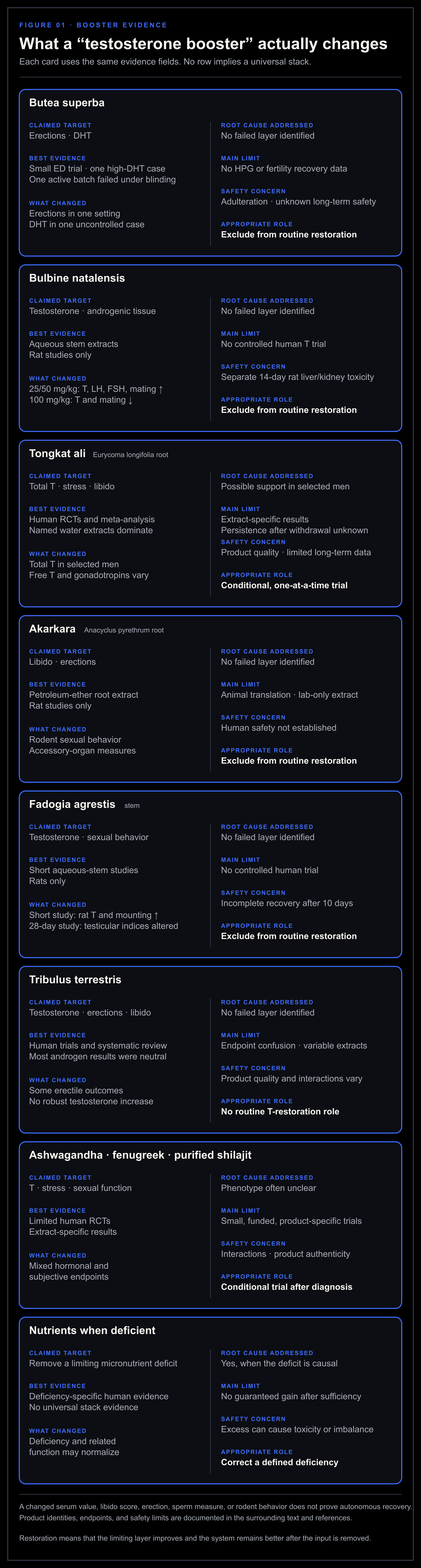
Figure 1. Recovery means that the product repairs the failing layer and that the improvement remains after the product is removed.
The test for a real restoration claim
A higher testosterone result can be temporary. Restoration requires the limiting layer to improve and the result to persist after withdrawal.
Before using a product or tactic, write down:
The symptom or laboratory pattern being treated.
The biological layer thought to be limiting.
The exact product, extract, or exposure.
The human evidence for that population and endpoint.
The result that will be measured.
The safety, interaction, and fertility risks.
The stopping rule.
What should remain improved after withdrawal.
When the limiting layer is unknown, the product is an experiment on the output.
Start here: find the limiting layer
Diagnosis locates the weak link. Begin with the symptoms that carry the most androgen information, then use the timeline and laboratory pattern to separate the causes. Fatigue, low mood, poor concentration, weight gain, and weak training progress remain useful context, but carry little diagnostic weight alone.
Start with four checks:
Define the change from your own baseline. Record the symptom, when it began, whether it is constant or intermittent, and how much it affects daily life.
Check whether the change fits androgen function. Loss of libido, fewer spontaneous or morning erections, reduced testicular volume, delayed puberty, infertility, hot flushes, loss of body hair, and reduced shaving frequency carry more androgen information than fatigue alone.[40][41]
Record what changed before the symptom. Review sleep, shift work, weight, food intake, training load, illness, pain, medication, alcohol, opioids, hormones, anabolic steroids, head injury, testicular injury, and fertility plans.
Choose urgent care, laboratory testing, or stabilization. Sudden severe testicular pain, a testicular mass, a severe new headache with visual change, or serious systemic illness bypasses self-directed optimization.
Define the problem before ordering a panel
Use three symptom groups.
Sexual and reproductive changes include libido, spontaneous erections, erectile reliability, ejaculation, testicular change, fertility, and breast symptoms. These are the most useful starting signals, though each still has non-androgen causes.
Physical and recovery changes include strength, muscle loss, bone injury, anemia, reduced work capacity, slow recovery, and a simultaneous fall in training performance. These become more informative when several change together.
General symptoms include fatigue, low mood, irritability, poor concentration, sleepiness, and reduced initiative. Keep them in the record. Do not let them carry the diagnosis alone.
For every symptom, record the direction of change, the date it began, and the context. A lifelong low libido is a different problem from a sharp decline after an anabolic-steroid cycle. Poor recovery during a severe cut is different from progressive weakness with a testicular mass.
Mark the context that changed
Build a simple twelve-month timeline. Include:
Sleep timing, snoring, witnessed breathing pauses, travel, and shift work.
Weight, waist, dieting, fasting, appetite, and food restriction.
Training volume, intensity, endurance work, injuries, and deloads.
Acute or chronic illness, pain, fever, gut symptoms, and major stress.
Every prescription, supplement, hormone, prohormone, selective androgen-receptor modulator, and recreational drug.
Head injury, testicular injury, infection, surgery, chemotherapy, and radiation.
Puberty, previous fertility, current fertility goal, and anabolic-steroid history.
The sequence often identifies the first branch. Symptoms that follow sleep fragmentation point toward timing and airway work. Symptoms that follow rapid weight loss and rising training load point toward low energy availability. Low testosterone after androgen use begins in the suppression branch. A long fertility history with small testes or high follicle-stimulating hormone begins in the testicular branch.
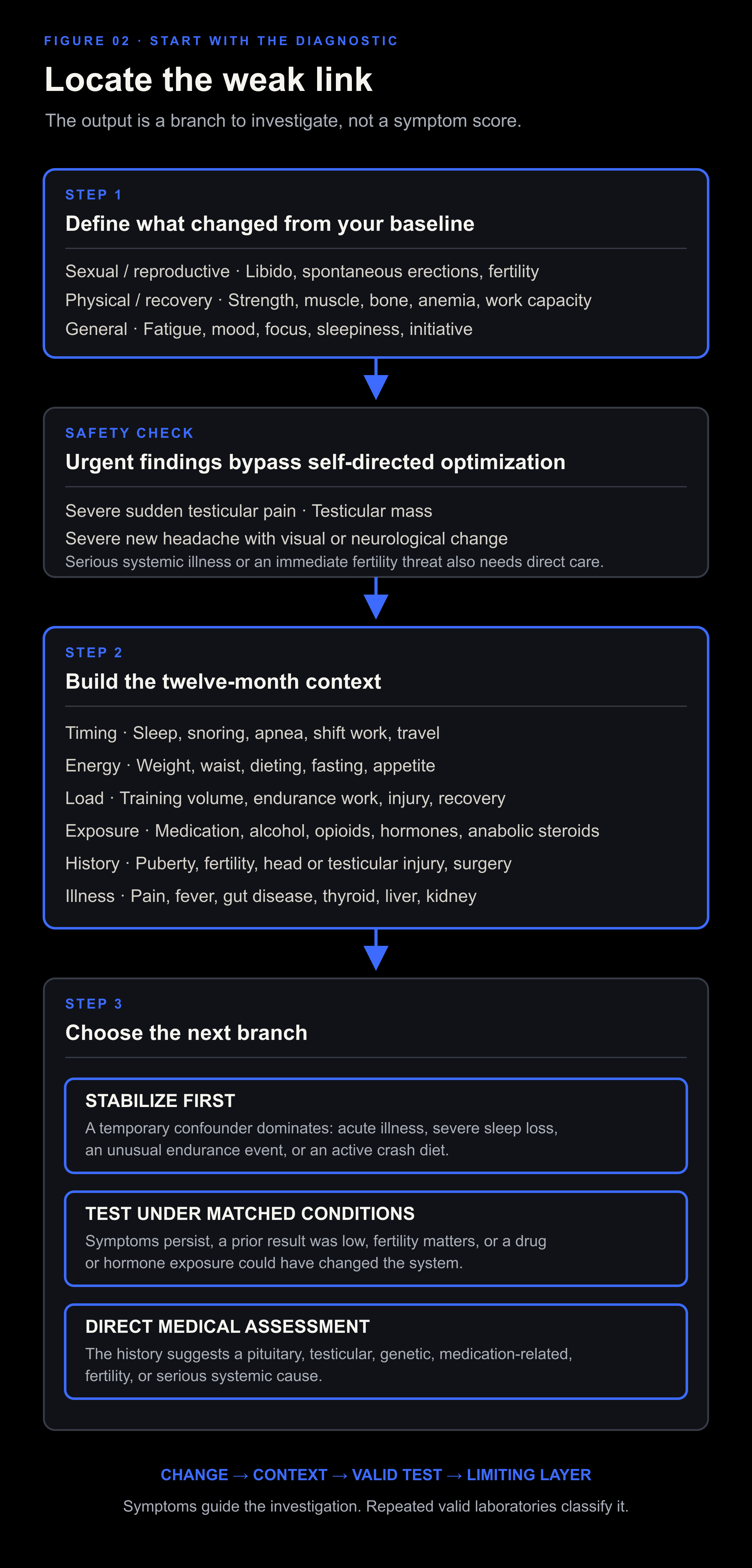
Figure 2. Define the change, screen urgent findings, record the context, and then choose stabilization, matched laboratory testing, or direct medical assessment. The figure is a decision sequence, not a symptom score.
Decide whether to stabilize, test, or escalate
Stabilize first when a temporary confounder clearly dominates and no red flag is present. Examples include acute febrile illness, several nights of severe sleep loss, an unusual endurance event, or an active crash diet. Correct the condition, allow recovery, and then test under representative conditions if symptoms remain.
Test now when sexual or reproductive symptoms persist, when several physical signs move together, when a previous result was low, when fertility is at risk, or when medication or hormone exposure could have suppressed the system.
Seek direct assessment when the history suggests a pituitary, testicular, genetic, medication-related, or serious systemic cause. Lifestyle work can continue when safe. It should not delay the cause-specific evaluation.
This first pass narrows the question. Laboratory testing later in the article separates low production, weak central signaling, testicular resistance, altered binding, exogenous suppression, and symptoms that are probably coming from another system.
The Testosterone Kabbalah
The Kabbalah is a map of dependencies. No knowledge of religious Kabbalah is needed to use it.
A vertical path runs through the middle. It starts with the conditions that let cells work, then follows cholesterol into the mitochondrion, through steroid production, into testosterone, and finally into tissue action. Side nodes show the systems that supply timing, fuel, instructions, transport, and clearance.
The map prevents a common mistake. A man can have enough cholesterol and still have weak LH signaling. He can have strong LH and damaged Leydig cells. He can have adequate testosterone in blood and poor sleep, vascular erectile dysfunction, low free testosterone, or impaired fertility. Every layer has to carry its part of the system. Raising one downstream number leaves failure elsewhere untouched.
How to read each part of the map
Timing comes first. Light reaching the retina sets the central circadian clock. Sleep supplies the cycles during which testosterone normally rises. An obstructed airway can fragment those cycles.
Food supplies energy and raw material. Protein, carbohydrate, fat, vitamins, and minerals support the tissues involved. The body can make cholesterol from carbon supplied by any macronutrient. High fat intake is unnecessary for that purpose.
Thyroid and mitochondria provide working capacity. Thyroid hormone helps regulate metabolic activity. Mitochondria are small structures inside cells that turn fuel into usable energy and host the first regulated step of steroid production.
The brain and pituitary provide instructions. The hypothalamus is a control region in the brain. It signals the pituitary gland, which releases LH and follicle-stimulating hormone, usually shortened to FSH. LH tells Leydig cells in the testes to make testosterone. FSH supports Sertoli cells and sperm production. Together, this is the hypothalamic-pituitary-gonadal axis, shortened to the HPG axis.
Cholesterol must reach the correct place. Steroidogenic acute regulatory protein, shortened to StAR, moves cholesterol to the inner mitochondrial membrane. The enzyme CYP11A1 then converts it into pregnenolone, the first steroid in the pathway.
Later enzymes finish production. Pregnenolone can pass through several intermediates before becoming testosterone. Enzyme activity, cellular energy, reducing power, and testicular health determine how much material moves through the pathway.
Blood transport changes the measured result. The liver makes SHBG, a protein that carries testosterone. Liver health and metabolic state can change SHBG and therefore change how total and free testosterone relate.
Tissues decide the final signal. 5-alpha-reductase converts testosterone to DHT. Aromatase converts it to estradiol. Receptors in each tissue respond to the hormones that reach them. The blood value cannot describe every local conversion.
Gut and liver remain supporting systems. The gut handles digestion, absorption, immune exposure, serotonin production, and microbial metabolites. The liver makes binding proteins and clears steroid hormones. These are defined functions rather than vague detox claims.
Body composition and suppressive exposures modify the network. Excess adipose tissue can lower SHBG, increase aromatase, worsen insulin resistance, and contribute to sleep apnea. Severe underfeeding can also suppress reproductive signaling. Illness, medication, previous androgen use, and training that exceeds recovery can interrupt several nodes at once.
The cobalt path in the figure marks production from cholesterol transport to tissue action. Gray lines show support or context. The smaller LH node represents the immediate testicular instruction. It is essential without being the whole system.
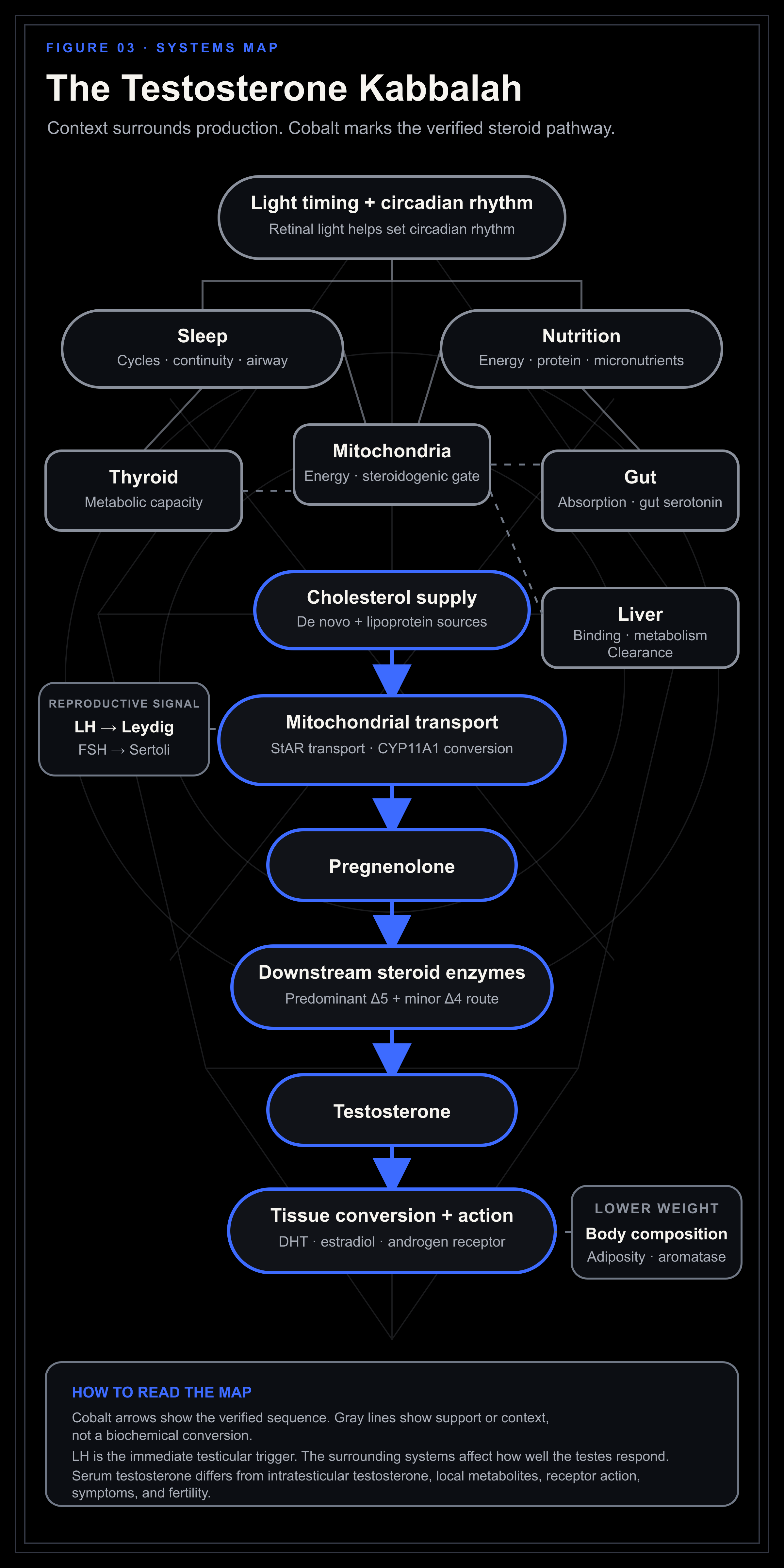
Figure 3. Cobalt follows the regulated steroid pathway. Gray marks timing, energy, metabolic support, transport, clearance, and lower-weight modifiers. The smaller reproductive node separates LH action on Leydig cells from FSH support of Sertoli cells.
Part I. How the system works
1. A testosterone result has several layers
Testosterone exists at several biological levels at once:
Leydig cells make it.
A blood sample measures it.
SHBG and albumin carry it.
Tissues convert it to DHT or estradiol.
The androgen receptor receives the signal.
The signal contributes to libido, fertility, blood production, bone, muscle, mood, and body composition.
Each layer can move independently.
Laboratory testosterone is one output. The final androgenic signal is the wider result. A higher blood value can still coexist with poor sleep, low energy availability, thyroid disease, inflammation, vascular erectile dysfunction, or weak tissue response.
Production, concentration, and action
Production means how much the testes secrete over time. Concentration means how much a laboratory finds in one sample. Action means what happens inside a tissue.
Concentration changes when production changes, but also when SHBG, clearance, plasma volume, sampling time, or the assay changes. Tissue action varies again with local conversion, receptor function, tissue health, and other hormones.
This explains several common mismatches:
Low total testosterone with low SHBG can coexist with a smaller change in free testosterone.
Normal total testosterone with high SHBG can coexist with low free testosterone.
Exogenous testosterone can produce a high serum concentration while suppressing LH, FSH, intratesticular testosterone, and sperm production.
Normal testosterone can coexist with erectile dysfunction from vascular disease, medication, neuropathy, anxiety, sleep loss, or relationship context.
Acute illness or underfeeding can lower testosterone without permanent testicular failure.
Aim for better function and a stable, healthy system. Do not chase the highest serum number.
2. Testosterone rises through sleep cycles
Circadian rhythm sets the clock. It is the body’s internal timing system, running on a cycle of roughly twenty-four hours. Outdoor light reaching the eyes is one of its main environmental setters.[42][43]
Sleep supplies the cycles. Testosterone starts rising after sleep begins. In healthy young men, it reaches a high level around the first rapid eye movement, or REM, period and stays elevated until waking.[44][45] Fragmentation can delay that rise. Selective loss of slow-wave sleep can also lower the morning result.[46][47]
Sleep deprivation lowers testosterone. The clearest adverse evidence comes from total sleep deprivation.[48] Severe repeated restriction has also lowered daytime testosterone in a controlled study.[49]
Cycles are not fixed ninety-minute blocks. Their length varies within the same person and across the night. Slow-wave sleep is usually concentrated earlier. REM periods become longer later. Cutting the last part of the night removes REM-rich sleep. Fragmenting the first part damages slow-wave sleep.
Protect complete cycles. Allow enough time for sleep and reduce avoidable awakenings. Judge the result through morning function, daytime alertness, symptoms, and matched laboratory tests.
Build the sleep window around enough complete cycles and a reasonably stable wake time. Shift work, travel, parenting, illness, and chronotype can move the clock. The standard remains adequate opportunity, intact cycles, regular timing, and alignment with daylight.
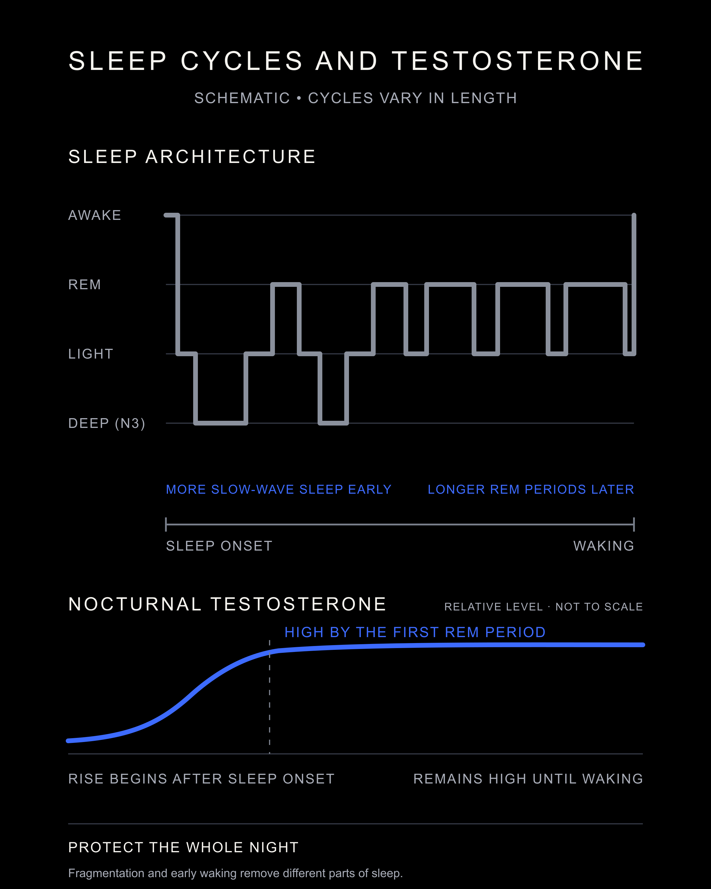
Figure 4. Testosterone starts rising after sleep onset. Slow-wave sleep is heavier early in the night. REM periods lengthen later. Fragmentation and a shortened night remove different parts of the cycle.
Circadian rhythm and testing
The circadian clock places sleep and hormone rhythms in time. Sleep provides the stages and continuity.
Light can shift the clock without opening an obstructed airway. Extending the sleep window cannot fully repair repeated night-shift misalignment.
A blood draw at 8:00 a.m. can mean different things in a day worker, a night worker, and a man who slept from 4:00 a.m. to noon. Record:
Wake time on the day of the test.
Sleep start and duration for the preceding nights.
Whether the schedule was typical.
Recent night shifts or travel across time zones.
Acute illness, severe training, fasting, or caloric restriction.
Time of the blood draw relative to waking.
In the HORMONIT night-work study, androgen and melatonin timing shifted later, while individual sex steroids followed different patterns.[50] The sampling plan has to account for that shift.
Pituitary hormones do not share one clock
Each pituitary hormone has its own timing.
LH is pulsatile. Researchers sampled twenty healthy men every ten minutes and found LH intervals ranging from 30 to 480 minutes. A daily LH pattern appeared across the group, while FSH showed no clear daily rhythm.[51] One isolated LH result may catch a pulse, a trough, or the interval between them.
Prolactin and growth hormone follow sleep closely. Thyroid-stimulating hormone, or TSH, reflects both sleep and circadian timing. Cortisol follows the circadian clock more strongly.[52]
In a small crossover study, ten young men followed a later sleep-and-meal schedule for three weeks. Free testosterone and prolactin fell, and several measured twenty-four-hour rhythms became much flatter.[53] The experiment moved sleep and meals together, so its result belongs to the shifted schedule as a whole.
Melatonin is also time-dependent. Retinal light remains the main environmental signal for the central clock. Melatonin can shift circadian phase, but the effect depends on timing. In one 3 mg study, doses taken during the first half of habitual sleep produced little phase shift.[54][55]
Melatonin changes one timing signal. The schedule, sleep pattern, symptoms, and matched laboratory results show whether the wider hormonal rhythm is working.
Sleep opportunity and common disruptors
Time in bed is not time asleep. A useful sleep window includes the sleep itself, the time needed to fall asleep, and ordinary brief awakenings.
Build from the wake time. Choose a workable wake time and count backward far enough to complete the night. When spending longer in bed only creates more wakefulness, persistent insomnia may require cognitive behavioral therapy for insomnia.
Caffeine depends on dose and timing. It can reduce total sleep time, sleep efficiency, and deep sleep.[25] Find the latest dose that leaves sleep unchanged, then leave some margin.
Alcohol can trade faster sleep onset for a worse second half of the night. It can also worsen sleep-disordered breathing.[56]
Pain, reflux, nocturia, nicotine, stimulants, heat, noise, and a partner’s schedule can all break sleep cycles. Find the main disruptor before reaching for a sleep supplement.
Restless legs syndrome deserves its own check. The pattern is an urge to move the legs that begins or worsens at rest, improves with movement, and is usually worse in the evening. Clinically important restless legs can delay sleep and cause repeated disruption. Review ferritin and transferrin saturation with a clinician, untreated apnea, alcohol, caffeine, and medicines that can aggravate the condition. This includes some antihistaminergic, serotonergic, and antidopaminergic drugs.[57] Iron is not a blind sleep supplement. Its use depends on the iron studies and the clinical pattern.
Sleep continuity and the airway
Enough time in bed will not compensate for an airway that keeps closing.
Look for loud snoring, witnessed pauses, gasping, dry mouth, morning headaches, nocturia, unrefreshing sleep, and daytime sleepiness. Obstructive sleep apnea, shortened to OSA, is associated with lower testosterone, especially when disease is severe.[58] Obesity contributes. Lean men can also have apnea through jaw structure, tonsils, or nasal obstruction.
Nasal allergies can fragment sleep. Pollen, dust mites, animal dander, mold, smoke, infection, reflux, and structural narrowing can all block the nose, so treatment should follow the cause.
Use this order:
Reduce the known allergen or irritant.
Use a saline rinse when it helps rhinitis. Use only distilled, sterile, or previously boiled and cooled water.[59][60]
Try a nasal strip when the nasal valve is narrow. It may improve airflow. It does not treat obstructive sleep apnea.[61]
Seek examination for persistent obstruction, recurrent sinus disease, enlarged tonsils, or a deviated septum.
Get a sleep study when apnea signs are present.
A true food allergy or reflux can worsen congestion in some people. Treat a suspected food as relevant when the reaction repeats under similar conditions. “Starch” is too broad to serve as a diagnosis.
Mouth-taping evidence is small, and obstruction or sleep apnea makes the practice unsafe.[62] Open the nose and rule out apnea first.
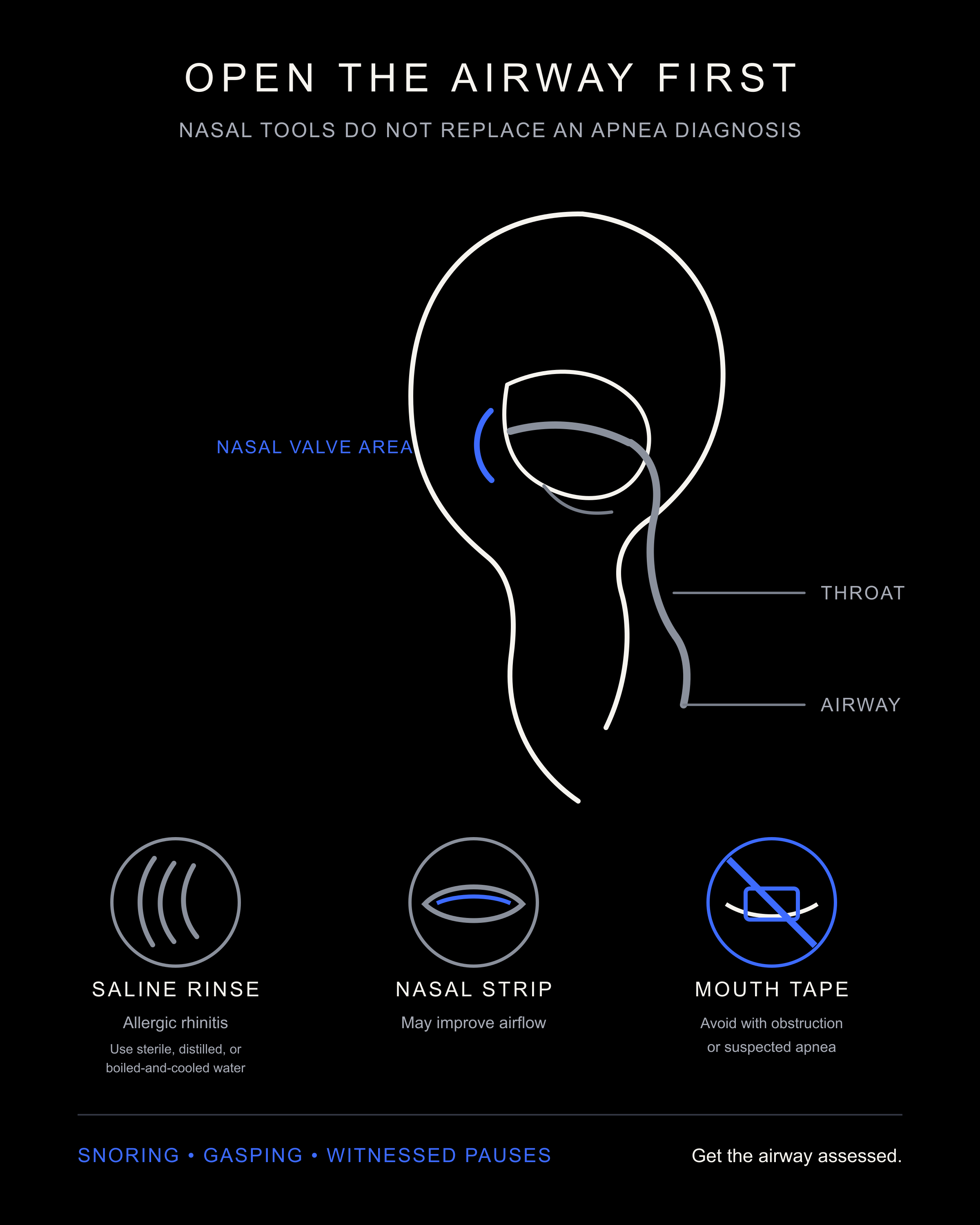
Figure 5. Rinse and nasal strips can help selected nasal problems. Mouth taping is not an apnea treatment and should never be used through an obstructed nose.
Continuous positive airway pressure, or CPAP, treats diagnosed apnea but does not reliably raise testosterone on its own.[63] Its purpose is to keep the airway open and improve breathing, oxygenation, and sleep quality.
3. Retinal light, dopamine, and blue light
Light reaching the retina is the main environmental signal for the central clock. Specialized retinal cells respond strongly to short wavelengths and send that timing information to the brain.[64]
You need thyroid, you need the amino acid tyrosine, and you need your retina to be exposed to light.
Tyrosine is a dopamine precursor, and tyrosine hydroxylase controls the rate-limiting step in catecholamine synthesis. Bright light can stimulate dopamine synthesis inside the retina.[65][66]
Outdoor light supports testosterone through more than one route. It anchors circadian rhythm, supports retinal dopamine, and protects sleep. In the hypothalamus, dopamine restrains pituitary prolactin. When prolactin remains pathologically high, GnRH and LH can fall, followed by testosterone, libido, erections, and fertility.[42][43][67][64][65]
Where bromantane fits
Bromantane, also called Ladasten, connects to this dopamine discussion through a different compartment. Outdoor light begins with photons at the retina. Bromantane acts inside central catecholamine pathways.
The rat response changed by brain region and time. In adult male rats given 50 mg/kg orally, tyrosine-hydroxylase messenger RNA in the ventral tegmental area peaked at 220% of control after 90 minutes. In the hypothalamus, the same transcript first fell to 66% at 30 minutes, then rose to 212% at 90 minutes. Hypothalamic tyrosine-hydroxylase protein was about twice the control level after two hours. In the striatum, the transcript fell to about 20% at 30 minutes and returned to control by two hours, while the protein did not rise. Nucleus-accumbens L-3,4-dihydroxyphenylalanine, or L-DOPA, and dopamine were about three times control at two hours. Hippocampal L-DOPA and dopamine showed a similar threefold peak at 90 minutes.[68]
A separate microdialysis experiment followed five freely moving male rats after 100 mg/kg orally. Extracellular dopamine in the dorsal striatum peaked at 553% of baseline after two hours and remained between 240% and 355% through eight hours. At eight hours, the dopamine metabolites 3,4-dihydroxyphenylacetic acid, or DOPAC, and homovanillic acid, or HVA, had fallen to 68% and 60% of baseline. Tetrodotoxin almost stopped spontaneous dopamine release and only partly suppressed the bromantane response, which points to more than one release mechanism.[69]
In the gene-expression experiment, a 588-gene whole-brain macroarray identified 15 initial candidates 90 minutes after 50 mg/kg orally. Quantitative PCR supported 12 changes. The largest reported shifts included carboxypeptidase H falling to 10% of control, synapsin I to 42%, gamma-aminobutyric acid transporter 3, or GAT3, to 60%, the APC tumor-suppressor transcript rising to 221%, cytoplasmic actin to 180%, and the retinoblastoma transcript, Rb, to 176%. The authors treated GAT3 and carboxypeptidase H as possible pharmacological targets. They treated several other changes as compensatory or neuroplastic responses.[70]
In rat hippocampal slices, 10 micromolar bromantane converted weak short-term potentiation into a long-lasting form. Blocking protein synthesis prevented the effect. A D1/D5 antagonist given during induction also prevented the conversion. Starting the antagonist 40 minutes after induction did not. This supports a dopamine- and protein-synthesis-dependent synaptic mechanism in that preparation.[68]
The two routes should be read separately:
Outdoor light: Retinal photoreception, circadian timing, and dopamine production inside the retina.
Bromantane: Pharmacological regulation of dopamine synthesis and signaling inside selected brain regions.
The androgen route is indirect. Hypothalamic dopamine restrains prolactin. Pathologically high prolactin can suppress GnRH, LH, testosterone, libido, erections, and fertility. These bromantane papers stop upstream. They contain no prolactin, GnRH, LH, testosterone, or retinal-dopamine measurement.[68][71]
Treat bromantane as central dopaminergic pharmacology. It cannot replace daytime outdoor light and it sits outside the default natural protocol. The milligram-per-kilogram values above describe rat experiments. They are not human dosing instructions. Bromantane is a CNS-active drug. Product identity, medication interactions, sleep timing, and the clinical context matter.
Daylight contains plenty of blue light, which supports alertness and circadian anchoring during the day. Exposure to the same spectrum at night can suppress melatonin and delay sleep timing.[42][43][64]
Use a strong day-night contrast:
Get outdoor light after waking when feasible.
Work in a bright environment during the day.
Dim the environment as sleep approaches.
Reduce bright screens near the face at night.
Keep the bedroom dark.
Someone trying to delay an unusually early clock may need morning light later. Clinicians may also use evening light deliberately for some circadian disorders. Timing determines whether light advances or delays the clock.
Calibrate.day turns wake time into a simple light and wind-down schedule. Use it to organize timing. Circadian phase measurement and sleep diagnosis belong to clinical assessment.
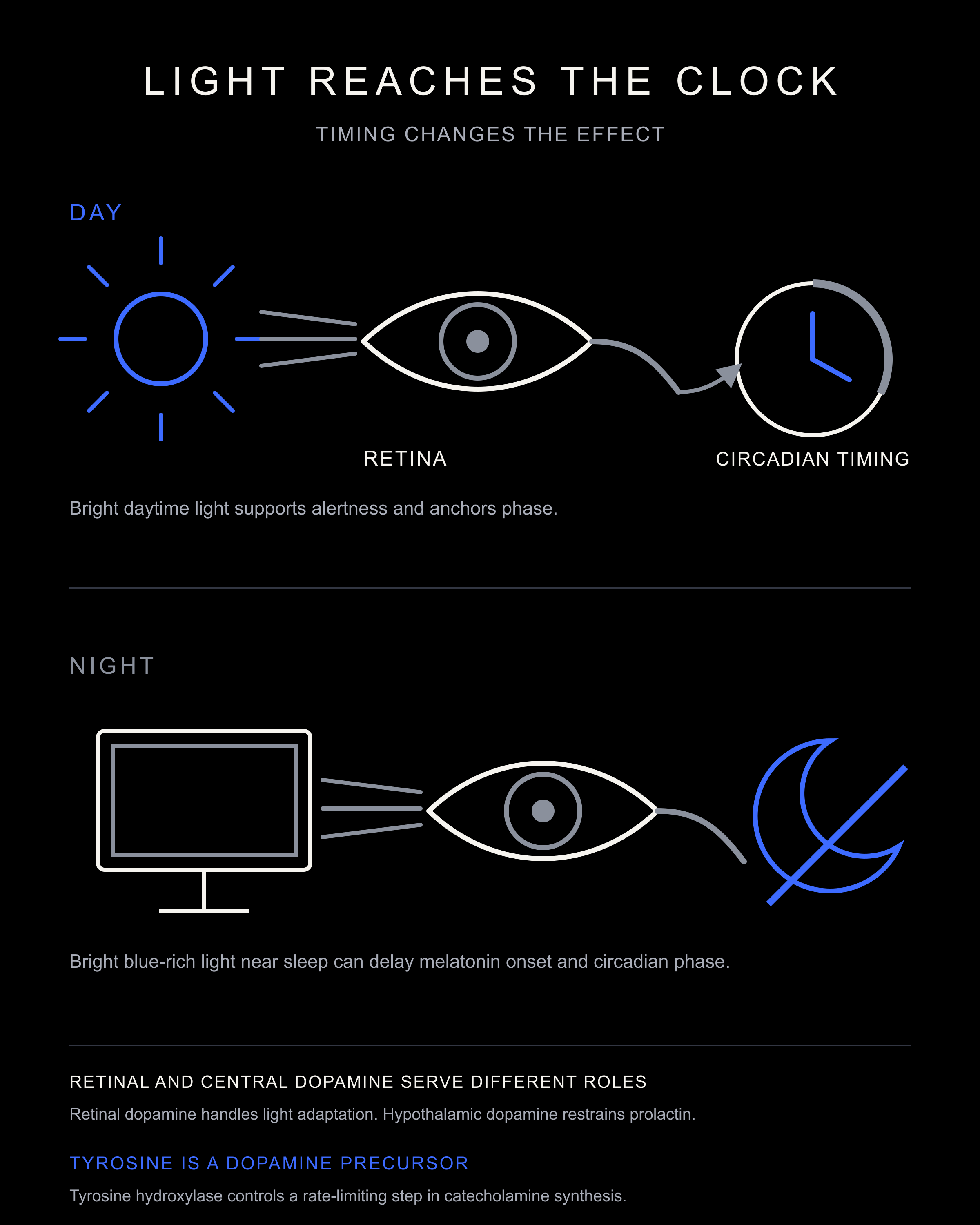
Figure 6. Daytime retinal light supports circadian timing and retinal dopamine. Hypothalamic dopamine affects the reproductive axis separately through prolactin control.
Retinal light and skin exposure act through different routes. Correct vitamin D deficiency for its established indications. In men who begin with normal testosterone, supplementation trials show no reliable testosterone increase.[21] Set skin exposure by latitude, season, skin type, and medical risk instead of a universal number of minutes.
Seasons change the comparison
Male hormone results can vary across the year. Two large observational datasets found seasonal variation in testosterone and placed the peak in different parts of the year.[72][73] Latitude, temperature, activity, sleep, illness, body composition, and laboratory timing all shape the comparison.
Record the season when comparing tests taken many months apart. Keep the laboratory, wake context, and sampling method as similar as possible. Treat season as a comparison variable, never as an explanation for a persistent low result with symptoms.
4. Energy availability
Reproductive output responds to food intake, expenditure, stored energy, illness, and stress. One man may need more food while another needs slow fat loss. The starting state determines the intervention.
Low energy availability
Energy availability means the food energy left for normal physiology after exercise. Male studies do not support one universal cutoff. Use the man’s weight trajectory, intake, training, recovery, symptoms, and hormone pattern.
During contest preparation, testosterone in one bodybuilding case study fell from 9.22 to 2.27 ng/mL and recovered after the competition.[74] The athlete had combined a long deficit, very low body fat, and high training demand.
Short trials have produced different results. In one randomized crossover, six exercising men completed four days at about 16 kcal/kg fat-free mass/day. Leptin and insulin fell, but testosterone and free triiodothyronine, or T3, did not.[75] Two other trials found lower testosterone at their lowest intake.[76][77] Severe restriction presents a real risk. The size and timing of the response vary, and a four-day experiment cannot define chronic adaptation.
Look for a pattern that includes several of the following:
Unplanned or rapid weight loss.
Persistent hunger or rigid food restriction.
Falling performance despite high effort.
Cold intolerance.
Loss of morning erections or libido.
Irritability, sleep disruption, or waking hungry.
Recurrent injury.
Training volume that rose while food intake stayed fixed.
A testosterone decline that followed a cutting phase.
Diagnose the pattern from weight change, intake, training, recovery, and the timing of symptoms. One isolated item carries little weight.
5. Nutrition, body composition, and the supplement reference
The evidence hierarchy for nutrition
Nutrition changes energy availability, body composition, training, sleep, liver function, thyroid function, and nutrient status. The strongest evidence supports three actions: correct a sustained deficit, reduce excess fat without crash dieting, and replace a genuine deficiency.
The trials do not identify one ideal macronutrient ratio. A 2021 analysis found lower testosterone with lower-fat diets, while a 2025 analysis that included more trials found no overall advantage for higher-fat diets.[78][79] Set the ratio from energy needs, digestion, training, body composition, and metabolic health.
Protein supports tissue repair and lean mass. Carbohydrate supports glycogen, training, and recovery. Fat supplies energy, essential fatty acids, and helps absorb fat-soluble nutrients. The useful amounts depend on the person.
Build the diet in this order:
Correct an energy deficit or excess that is impairing health.
Establish adequate complete protein.
Supply carbohydrate and fat in amounts that support training, sleep, glucose regulation, and the intended weight trajectory.
Build micronutrient sufficiency from tolerated foods.
Add a supplement only when the reason, dose, expected result, and stopping rule are clear.
The original Ray Peat sample food pyramid
The diagram below keeps the original labels and placement from the widely circulated sample pyramid. I have not renamed its tiers or added foods. The image itself says that these are not structured guidelines, and it remains a third-party summary of Ray Peat’s diet rather than a diagram published by Ray Peat.
Inside the pyramid are fruit, fruit juice, honey, dairy, eggs, collagenous meats, non-PUFA seafood, coconut oil, butter, daily carrots, and weekly liver. PUFA sits outside it. Ray Peat’s own writing supports many of these choices,[80][81][82] though none becomes a proven testosterone intervention by appearing in the image.
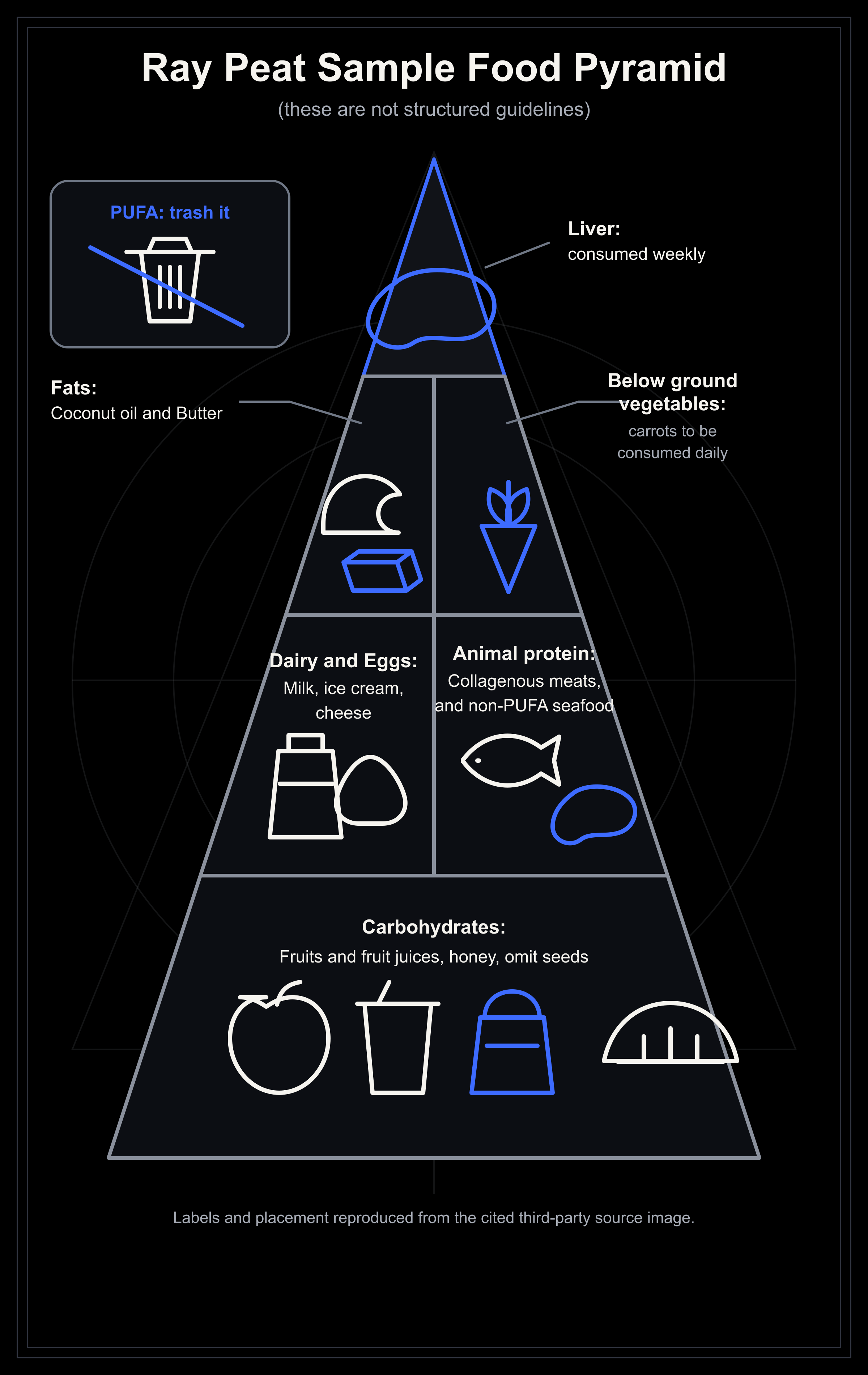
Figure 7. The labels and placement reproduce the original sample pyramid. “These are not structured guidelines” is part of the source image. The pyramid is a third-party summary, not a diagram published by Ray Peat.
Energy context
Carbohydrates occupy the base of the pyramid. The person’s energy state determines the amount.
A lean athlete who loses weight, morning erections, performance, and warmth during a hard cut may need more food and less training. A sedentary man with visceral fat, insulin resistance, low SHBG, and apnea may need gradual fat loss. Both patterns can lower testosterone through different routes.[74][83][84]
Use regular meals when long gaps lead to under-eating, waking hunger, poor training, or overeating. Meal frequency serves energy stability and adherence rather than a universal testosterone target.
Carbohydrate: source, amount, and purpose
Ray Peat favored ripe fruit, fruit juice, honey, and easily digested carbohydrates. Juice can be useful when appetite is low or expenditure is high. Whole fruit provides more fiber and chewing. Potatoes, rice, and other starches also belong when they improve satiety, digestion, or adherence.
Evaluate the carbohydrate pattern through:
Stable training and recovery.
Sleep without hunger or reflux.
Good digestion.
The intended weight and waist trend.
Appropriate glucose control when metabolic disease is present.
For testosterone, current human evidence does not rank fruit sugar above a tolerated, energy-matched starch. Choose the source by digestion, sleep, training, glucose control, and body composition.
Protein: complete sources and complementary gelatin
Choose complete protein sources that digest well. Ray Peat often favored milk, cheese, eggs, shellfish, low-fat fish, and ruminant meat, with liver in a smaller role.
Gelatin or collagen supplies glycine and can complement a diet heavy in muscle meat,[82] but it cannot replace complete protein.
Shellfish supply zinc, copper, and selenium. Eggs supply protein and choline. Liver is dense in vitamin A, copper, B vitamins, and iron, so the amount has to account for the rest of the diet.
A workable protein pattern has five features:
Intake supports lean mass, training, and recovery.
The diet remains energetically adequate.
Protein selection does not create persistent digestive symptoms.
Kidney, liver, allergy, or metabolic conditions are respected.
The plan does not depend on one food that the person cannot tolerate.
Dietary fat, cholesterol, and PUFA
Polyunsaturated fatty acids are shortened to PUFA. The first article favored saturated fats and strict PUFA avoidance. Ray Peat preferred lower-PUFA foods and stable cooking fats,[81] while the human testosterone evidence supports a much narrower claim.
Some studies report lower testosterone with very low fat intake. A newer analysis of trials found no overall advantage for higher-fat diets.[78][79]
The body can make cholesterol from carbon supplied by carbohydrate, fat, and protein. Dietary cholesterol contributes to that pool, alongside cholesterol made inside the body, circulating lipoproteins, lipid droplets, and cell membranes.[85][86][87][88] Steroid production does not require forcing fat intake upward.
Four constraints matter when setting fat intake:
Keep total fat high enough for nutritional adequacy and food tolerance.
Keep total energy aligned with the intended weight trajectory.
Minimize repeatedly heated or oxidized oils and the ultra-processed foods that carry them.
Choose cooking fats and whole-food fat sources that fit cardiometabolic risk, digestion, and preference.
The source pyramid names coconut oil and butter, while my first article also named dairy fat, cocoa butter, and ruminant fat. They can be used when they fit the person and the diet. Keep PUFA low without chasing literal zero. Complete elimination is impractical.
Minerals, vitamins, and dose
The first article named calcium, salt, magnesium, thiamine, niacinamide, riboflavin, biotin, vitamin K2, vitamin E, zinc, selenium, iodine, choline, taurine, glycine, and creatine. Separate them into three categories.
Required nutrients. Calcium, magnesium, potassium, sodium, iodine, selenium, zinc, B vitamins, and fat-soluble vitamins serve established physiological functions. An adequate diet should cover them because deficiency can impair general and reproductive health.
Context-dependent additions. Creatine can support repeated high-intensity performance. Gelatin or glycine can complement the protein pattern. Choline-rich foods contribute to ordinary nutrient sufficiency. Coffee can remain in the diet when caffeine leaves sleep intact. None guarantees a rise in testosterone.
Dose-sensitive compounds. Iodine, selenium, zinc, vitamin A, vitamin E, niacinamide, and salt can cause harm when the dose exceeds the person’s need or conflicts with disease or medication. Salt intake has to reflect sweat loss, climate, blood pressure, kidney function, and treatment context. Caffeine timing has to protect sleep.[25]
Salt and the stress-hormone loop
Salt acts upstream of testosterone by changing the amount of hormonal compensation required to maintain circulation and sodium balance.
When sodium intake or effective blood volume falls, the kidneys release renin. Renin drives angiotensin II and aldosterone. Aldosterone conserves sodium, while sympathetic activity helps defend pressure and circulation. Across controlled diet studies, lower sodium increased renin, aldosterone, noradrenaline, and adrenaline on average.[89]
Ray Peat placed serotonin and prolactin inside the same loop. Sodium restriction can increase serotonin and sympathetic activity. Serotonin can increase prolactin. Prolactin can promote urinary sodium loss. Continued sodium loss then creates more demand for renin, aldosterone, and adrenaline.[90]
This explains the anti-prolactin role of adequate salt. Salt does not block the prolactin receptor. It reduces one of the conditions that can sustain serotonin, prolactin, and stress-hormone compensation. Ray Peat also connected sodium with sodium-potassium ATPase activity, fuel use, carbon-dioxide production, heat, and sleep. In this model, adequate salt supports the energetic state that keeps the stress system quiet.[90]
Cortisol needs precise wording. In a 630-person crossover intervention, liberal sodium increased 24-hour urinary free cortisol while producing a small decrease in morning serum cortisol. More cortisol in urine can reflect greater excretion rather than greater exposure inside tissues.[91] A single urinary or morning value cannot describe the whole cortisol rhythm.
Salt also sits beside thyroid, progesterone, and estrogen in this model. Progesterone opposes aldosterone signaling. Hypothyroidism and a high estrogen-to-progesterone state can favor sodium loss. Salt supports the resulting physiology without acting as a direct anti-estrogen drug.[90]
Use appetite, food intake, sweat loss, climate, blood pressure, kidney function, heart function, and medication together. A man losing sodium through heat, endurance work, diarrhea, or a low-salt diet has a different problem from a man with salt-sensitive hypertension, edema, kidney disease, heart failure, or a drug that changes sodium handling.
Thiamine and the energy layer
Thiamine becomes thiamine diphosphate, a cofactor for pyruvate dehydrogenase, alpha-ketoglutarate dehydrogenase, branched-chain alpha-ketoacid dehydrogenase, and transketolase.[92] That places vitamin B1 between food and usable energy.
Pyruvate dehydrogenase converts pyruvate into acetyl-CoA. Alpha-ketoglutarate dehydrogenase keeps the citric-acid cycle moving. Together they let carbohydrate support adenosine triphosphate, or ATP, production and carbon dioxide. Steroidogenic cells need ATP and reducing power to transport cholesterol and run steroid enzymes. When thiamine-dependent metabolism is constrained, the same LH signal reaches a Leydig cell with less energetic capacity.
The dopamine link follows the same logic. Tyrosine and vitamin B6 sit closer to dopamine synthesis, while thiamine supports the energy, redox state, and mitochondrial function needed by dopaminergic neurons. Thiamine supports the terrain in which dopamine is made and released. Dopamine agonists act directly at dopamine receptors. Thiamine works through metabolism.
The gut adds another route. Enteric nerves and intestinal smooth muscle require ATP. Pyruvate-derived acetyl-CoA also supplies the acetyl group used to make acetylcholine, a major signal for intestinal movement. A 2026 genetic study of 268,606 people linked stool frequency with vitamin B1 transport and metabolism. Dietary thiamine intake tracked with stool frequency in the accompanying nutritional analysis.[93] Better transit can reduce prolonged fermentation, constipation, and the gut burden that feeds endotoxin and peripheral serotonin signaling. Georgi Dinkov had already placed thiamine in this gut-motility context.[94]
Georgi Dinkov groups thiamine, niacinamide, and biotin around glucose oxidation.[95] A separate Georgi Dinkov article on testicular energy metabolism places aspirin and niacinamide in the same direction.[96] Together, these inputs aim at parts of the downstream metabolic state associated with T3: stronger glucose oxidation, better mitochondrial throughput, and less reliance on stress metabolism. T3 still has its own receptor-mediated actions.
Thiamine acts upstream of testosterone. It removes an energetic bottleneck when intake, absorption, transport, or enzyme activity is limiting. Once those enzymes are working normally, adding more B1 is a different intervention. Gram doses are pharmacological and should not be confused with ordinary nutritional sufficiency.
Zinc shows why context matters. A small 1996 study induced marginal zinc deficiency in four young men and supplemented nine marginally deficient older men. Testosterone fell during restriction and rose during repletion.[1] The result supports correcting deficiency, not giving high-dose zinc to men who already have enough.
Use the same rule here. Food history, clinical risk, and laboratory assessment can identify a real deficiency. Correct one defined problem at a time.
The supplement map: match the tool to the layer
A useful supplement has a named job. It corrects a shortage, treats a defined condition, or improves a measured function that is limiting the network. The name on the bottle is not enough. Form, dose, route, strain, timing, starting state, and endpoint decide what the evidence means.
Use this section as a reference by limiting layer. The ordered protocol in Part III determines whether any item belongs in the plan.
Use five evidence classes:
Deficiency correction. A nutrient restores a function that low intake, poor absorption, loss, or disease had constrained.
Condition-specific treatment. A product helps a defined phenotype such as antibiotic-associated diarrhea, irritable bowel syndrome with diarrhea, or documented male-factor infertility.
Functional support. A product improves sleep, bowel function, training performance, or another upstream input without directly raising testosterone.
Preliminary hormone signal. A small or uncontrolled human study moves a hormone but does not establish durable restoration.
Mechanism only. A cell or animal experiment explains a possible route. It does not establish the same outcome in men.
Every supplement enters through one layer. Judge it by that layer before judging testosterone.
Layer 1. Circadian rhythm and sleep
Glycine can support sleep and improve the amino-acid balance of a diet heavy in muscle meat. In a small crossover study of people dissatisfied with their sleep, 3 grams before bed improved subjective sleep quality and shortened the latency to sleep and slow-wave sleep. It did not change the proportion of sleep spent in each stage.[97] Glycine can be tested when it is tolerated. It does not repair apnea, nasal obstruction, pain, late bright light, or circadian misalignment.
L-theanine is another optional sleep aid. A recent systematic review found small improvements in selected subjective sleep outcomes across a mixed literature. Evidence for pure L-theanine was thinner than the marketing suggests.[98] It can support a wind-down routine when it is tolerated. It cannot replace light timing, complete cycles, or treatment of an airway problem.
Gelatin and collagen supply glycine. They can complement muscle meat and increase total glycine intake. Their effect depends on the amount actually consumed. They do not replace complete protein.
Melatonin can shift circadian timing and help selected sleep problems when timing and dose fit the problem.[99] It cannot replace daytime light, darkness at night, complete sleep cycles, or an open airway.
Epithalon, also called Epitalon, is the experimental tetrapeptide AEDG. Its relevant placement is the circadian branch. An experiment in old female rhesus monkeys changed the evening melatonin response and the cortisol rhythm.[100] A recent review describes a wider experimental literature.[101]
My overall response to Epithalon was positive. I did not preserve the preparation, route, dose, timing, cycle length, concurrent inputs, or raw sleep measurements.
Other public reports around Epithalon repeatedly mention deeper sleep, vivid dreams, clearer mornings, and easier circadian adjustment. Some instead describe transient sleep disruption, paradoxical stimulation, or difficulty waking. These reports identify possible response patterns. They provide no estimate of response frequency. Baseline state, light timing, concurrent drugs, preparation, dose, and product identity remain unresolved.
The practical route is circadian. Better melatonin and cortisol timing can improve sleep continuity, morning energy, metabolic control, and the nocturnal environment in which testosterone production occurs. Judge Epithalon by sleep timing, ease of waking, body temperature, next-day energy, and measured rhythm when available. Its placement here is circadian support, not direct LH stimulation.
Epithalon stays outside the default natural restoration plan until a specific question, a credible preparation, medical oversight, measurable endpoints, and a stopping rule justify the experiment.
Layer 2. Thyroid production and mineral sufficiency
Iodine is a raw material for T4 and T3. Selenium is built into the deiodinases that activate and inactivate thyroid hormone. Iron supports thyroid peroxidase. Zinc and vitamin A also participate in thyroid physiology. A systematic review of 57 human studies found that thyroid and iodine status are influenced by this wider micronutrient setting, while the randomized evidence did not support treating every association with more supplementation.[102]
The causal order is simple. An iodine shortage can limit hormone production. A selenium shortage can limit deiodinase function. Correcting either shortage can remove that bottleneck. Once iodine supply and selenoprotein activity are adequate, adding more does not keep pushing the thyroid forward.
Iodine has a narrow practical role. Review iodized salt, dairy, eggs, seafood, seaweed, thyroid disease, thyroid antibodies, and drugs that alter thyroid function. Concentrated kelp and iodine drops can move intake far beyond food-level exposure. Excess iodine can trigger hypo- or hyperthyroid patterns, especially in susceptible thyroids.[103]
Selenium needs the same restraint. Food intake, geography, malabsorption, thyroid diagnosis, and total supplemental exposure matter. In a 412-person randomized trial of autoimmune hypothyroidism, selenium and placebo produced equal improvement in quality of life.[104] Selenium is essential. Routine high-dose selenium is a different claim.
Myo-inositol becomes relevant here when the target is thyroid responsiveness. It supplies substrate to phosphoinositide signaling used downstream of TSH-receptor activation. In a randomized double-blind study, 46 women with autoimmune subclinical hypothyroidism completed six months of treatment. TSH fell from 4.4 to 3.1 mIU/mL with 600 milligrams of myo-inositol plus 83 micrograms of selenomethionine. TSH did not change with selenomethionine alone.[105]
The lower TSH is best read as reduced compensatory demand. When the thyroid responds better, the pituitary can maintain the system with less stimulation. The finding applies to myo-inositol plus selenium in women with autoimmune subclinical hypothyroidism. LH and FSH still require their own paired target hormones and clinical context.
Iron matters here only when deficiency is documented. Ferritin must be read with inflammation, transferrin saturation, blood count, symptoms, bleeding, and iron-overload risk. Blind iron can create a new oxidative and infectious burden.
Lactoferrin sits beside iron because it binds the mineral and can change its handling instead of simply delivering a large elemental dose. In a trial of 80 children with inflammatory bowel disease and iron-deficiency anemia, 100 milligrams per day for three months improved hemoglobin, serum iron, transferrin saturation, and ferritin. Interleukin-6, or IL-6, and hepcidin also fell. The response was stronger and better tolerated than ferrous sulfate in that population.[106] Lactoferrin is not a replacement for iron therapy in every cause of anemia.
Apolactoferrin is lactoferrin with very little iron attached. Its low iron saturation leaves more iron-binding capacity unoccupied. Native lactoferrin, apolactoferrin, and iron-saturated hololactoferrin are not the same preparation. Their iron saturation changes their physical behavior and can change the purpose of the product.[107] Check the label for iron saturation, dose, and intended target instead of treating every lactoferrin product as interchangeable.
Copper also requires context, especially after prolonged high-dose zinc.
Layer 3. Mitochondrial energy and recovery
Magnesium supports ATP handling, nerve and muscle function, glucose metabolism, and many enzyme systems. The first question is the amount of elemental magnesium. The second is intestinal tolerance. The salt attached to it matters, but it does not create a special testosterone pathway.
Magnesium citrate is well absorbed and can loosen stool. In a randomized crossover study in healthy men, citrate produced greater serum and urinary magnesium responses than oxide.[108]
Magnesium oxide supplies more elemental magnesium by weight and works as an osmotic laxative. Its absorption is lower than citrate in direct comparisons. It can still be the right form when constipation is part of the problem.
Magnesium glycinate or bisglycinate is often chosen when citrate is too laxative. A glycinate label does not prove a separate testosterone effect.
Magnesium chloride, lactate, gluconate, and aspartate are soluble oral forms. Choose among them by elemental dose, tolerance, cost, and the clinical use behind the product.
Magnesium malate, taurate, orotate, and L-threonate attach magnesium to different carriers. Claims about energy, the heart, or the brain require evidence for the complete compound. They cannot inherit a testosterone claim from magnesium itself.
Magnesium carbonate and hydroxide are used mainly in antacid or laxative products. Their purpose and elemental dose should be read from the complete label.
Magnesium sulfate is mainly a medical or laxative form. Intravenous magnesium is a drug treatment for specific conditions.
Topical magnesium chloride oil, creams, sprays, flakes, and Epsom-salt baths can feel more noticeable than an oral capsule. That practical response matters. A person may prefer the topical route for local muscle tension, cramping, relaxation, or sleep. The amount that crosses intact skin is not predictable from the product label or the sensation, and the human absorption literature remains limited.[109]
Correcting magnesium deficiency can remove a sleep or neuromuscular constraint. Across the sleep literature, observational associations are common and randomized results remain inconsistent.[110] Choose oral, topical, or bowel-active forms by the target and tolerance. When systemic deficiency is suspected, follow the intake, losses, symptoms, and laboratory context rather than estimating absorption from how strongly a product feels.
Kidney function sets the safety boundary. Reduced renal clearance can turn supplemental magnesium or magnesium-based laxatives into hypermagnesemia.[111]
Thiamine, riboflavin, niacinamide, magnesium, and lipoate support different steps in carbohydrate oxidation and mitochondrial redox handling. Biotin supports carboxylase reactions. Vitamin K2 as MK-4 has separate electron-carrier and steroidogenic hypotheses, but its testosterone evidence remains preclinical.[112] These compounds belong together only at the level of energy production. A shortage in one cannot be diagnosed from a testosterone result alone.
Taurine participates in bile-acid conjugation, osmoregulation, calcium handling, and neural signaling.[113] It was part of Georgi Dinkov’s one-month experiment. That combined self-experiment cannot isolate taurine as the reason testosterone rose.
Creatine monohydrate supports repeated high-intensity work and training quality. Its role here is performance and recovery. One small study changed the DHT-to-testosterone relationship, but that result has not made creatine a dependable DHT or testosterone treatment.[29]
Pinealon is the experimental tripeptide EDR. Its primary placement is brain metabolism, gene regulation, neuronal stress, and neurodegenerative models.[114] My overall response was positive, but I did not preserve the preparation, route, dose, timing, cycle length, concurrent inputs, or raw measurements. Reports of changed sleep or dreams can follow a change in brain function. They do not make Pinealon a sleep supplement or a sedative.
Pinealon fits under brain metabolism. It stays outside the default natural restoration plan until a specific question, a credible preparation, medical oversight, measurable endpoints, and a stopping rule justify the experiment.
Methylene blue is different. It is a drug-like redox compound, not another B vitamin. In experimental systems it can accept electrons from NADH and pass them to cytochrome c, creating an alternative route around blocked parts of the respiratory chain.[115] That mechanism explains why it appears in mitochondrial protocols. It does not establish a routine testosterone dose or a general need.
Methylene blue also inhibits monoamine oxidase A. Combining it with serotonergic drugs can cause serotonin toxicity.[116] Product purity, dose, medication interactions, and diagnosis decide whether it belongs anywhere. It stays outside the default natural protocol.
Layer 4. Prolactin, estrogen, and steroid signaling
Zinc can restore testosterone when zinc deficiency is the limiting problem.[1] It also supports spermatogenesis, immune function, and many enzymes. In a small acute experiment, oral zinc lowered basal prolactin in healthy adults.[117] That result supplies a direct anti-prolactin route. It does not show that chronic high-dose zinc treats persistent hyperprolactinemia. Chronic high doses can lower copper status. Read zinc beside diet, copper exposure, blood count, gastrointestinal disease, and the reason it is being used.
Vitamin B6 and its active coenzyme form, pyridoxal 5′-phosphate, or P5P, support amino-acid metabolism. Aromatic amino-acid decarboxylase uses P5P to make dopamine from L-DOPA. The same enzyme also makes serotonin from 5-hydroxytryptophan.[118] P5P therefore supports the dopamine route that restrains prolactin, but it is not a selective dopamine switch. Pyridoxine blunted the prolactin rise caused by exercise in one experiment, and a very small series of selected women with hyperprolactinemia reported responses.[119][120] A separate intravenous study in nine patients with hyperprolactinemia found no prolactin reduction.[121] Correct inadequate B6 and use the route when the clinical setting fits. Persistent hyperprolactinemia still requires diagnosis. Repeated high supplemental exposure can cause sensory neuropathy.[122]
Vitamin A supports epithelial differentiation and retinoic-acid signaling in spermatogenesis.[123] Liver, eggs, dairy, and carotenoid-rich foods contribute to status in different forms. Retinol is fat soluble and high supplemental doses can injure the liver.[124]
Vitamin D matters for bone, mineral handling, immune function, and deficiency correction. A randomized trial in healthy men did not produce a reliable testosterone increase.[21] Correct deficiency for its own reasons. Do not make testosterone the promise.
Vitamin E protects susceptible membrane lipids from oxidation. Ray Peat placed it more broadly as an estrogen antagonist because lipid peroxidation, inflammation, prostaglandins, and estrogen signaling can reinforce one another.[125] In one eight-week trial in 24 men receiving hemodialysis, vitamin E lowered prolactin but did not change free testosterone, LH, or FSH.[126] This supports an oxidative-stress and prolactin route in uremia. The wider anti-estrogen model is mechanistically coherent, but the dose and starting state still matter. Mixed tocopherols are not interchangeable with one isolated tocopherol, and very high doses can impair platelet function, especially beside aspirin.[24]
Vitamin K supports normal coagulation and bone-related proteins. For an androgen-focused K2 supplement, I prefer MK-4. MK-4 raised testosterone in rats and testis-derived tumor cells.[112] In contrast, an eight-week trial of 90 micrograms of MK-7 in women with PCOS found lower DHT relative to placebo, with no difference in total testosterone.[127] The trial did not measure 5-alpha-reductase itself, so the mechanism remains unresolved. The direction of the DHT result is enough for me to keep MK-7 out of a DHT-focused protocol. MK-4 is the preferred form here, while its direct testosterone evidence remains preclinical.
The other B vitamins remain part of the energy and blood-forming layers. Riboflavin, niacinamide, biotin, folate, and B12 should be corrected when diet, disease, medication, or measurement shows a reason. High-dose niacinamide is pharmacology, and high-dose biotin can distort thyroid, gonadotropin, prolactin, vitamin D, and other immunoassays that use biotin-based detection.[128] Tell the laboratory about every supplement before endocrine testing.
Layer 5. Gut barrier, motility, serotonin, and endotoxin
Zinc carnosine, enteric lactoferrin, riboflavin, and butyrate address different parts of this layer.
Zinc L-carnosine is a defined compound used for mucosal protection. In a small randomized crossover experiment, 10 healthy volunteers took 37.5 milligrams twice daily before indomethacin exposure. Zinc L-carnosine prevented the increase in small-bowel permeability seen with indomethacin alone.[129] That is a direct intestinal-barrier result. It is not the same intervention as ordinary zinc, and it does not show a testosterone effect.
Layer 2 covered lactoferrin as an iron-regulating protein. Its gut use raises a different issue: formulation. Enteric coating protects more of the protein from early gastric digestion and delivers it farther into the intestine. In one small eight-week trial of 26 adults with abdominal obesity, 300 milligrams per day of enteric-coated bovine lactoferrin reduced visceral-fat area more than placebo.[130] The study was too small to make lactoferrin a general fat-loss or testosterone product. Ordinary powders and non-enteric capsules cannot borrow an enteric-coated result.
Use enteric-coated lactoferrin when intestinal delivery is the target. Use an iron-defined lactoferrin or apolactoferrin preparation when iron handling is the target. The label must identify which intervention is actually being used.
Riboflavin, or vitamin B2, connects the gut and mitochondrial branches. In the randomized RIBOGUT trial, 50 or 100 milligrams per day for two weeks increased fecal butyrate without a gross change in microbial composition.[131] Riboflavin therefore offers one route for supporting microbial redox exchange and butyrate production. The trial did not test testosterone.
Butyrate is a short-chain fatty acid normally produced when colonic bacteria ferment suitable carbohydrate and fiber. Colon cells use it as fuel. Oral sodium butyrate is a delivery tool when endogenous production or colonic exposure is the target. Microencapsulation matters because free butyrate is absorbed early and has a strong odor and taste.
In a 66-person randomized trial, microencapsulated sodium butyrate added to standard IBS treatment reduced the frequency of selected symptoms. It did not produce a broad reduction in symptom severity.[132] Use sodium butyrate for a defined bowel target, such as an irritable-bowel-syndrome symptom pattern. It has no direct testosterone role.
Probiotics are strain-specific.
A bacterial species name is not a product specification. The strain, viable dose, formulation, storage, duration, and host condition are part of the intervention.
Saccharomyces boulardii is a probiotic yeast. CNCM I-745 is a named clinical strain and must remain on the product record when that is what was used. Across preparations identified as S. boulardii, a meta-analysis of 21 randomized trials found that antibiotic-associated diarrhea fell from 18.7 percent to 8.5 percent.[133] Antibiotics do not kill this yeast, while antifungal drugs can. It is inappropriate for critically ill or severely immunocompromised patients and for people with central venous catheters because fungemia has been reported.[134]
Heyndrickxia coagulans GBI-30, 6086, commonly still labeled Bacillus coagulans GBI-30, 6086 or BC30, has human evidence for bowel function. In a randomized trial of 111 adults with functional gastrointestinal complaints, one billion viable cells per day for four weeks improved selected stool and constipation outcomes.[135] The strain and viable count are part of the result. It did not test testosterone.
Limosilactobacillus reuteri ATCC PTA 6475 is the strain behind the famous testosterone claim. It preserved testicular size and testosterone in mice through an immune pathway.[136] A twelve-week double-blind trial then tested the same strain in healthy men aged 55 to 65. Neither the low nor high dose raised testosterone.[137] A later ten-person open-label pilot also found no meaningful change in sex hormones after twelve weeks, although body-fat, blood-pressure, and inflammatory measures moved.[138] The human endocrine results do not reproduce the mouse testosterone result.
Other L. reuteri strains have other jobs. BM36301 preserved testicular and endocrine measures in aged mice, but it has no comparable human testosterone trial.[139] DSM 17938 improved selected symptoms and bowel habit in a randomized adult functional-constipation trial.[140] NCIMB 30242 is a bile-salt-hydrolase-active strain that lowered LDL cholesterol in a trial of hypercholesterolemic adults.[141] Those results do not transfer between strains. A mixed product that merely says L. reuteri has not identified the intervention.
Many other L. reuteri strains exist. Each requires its own human endpoint. Species-level marketing cannot fill in missing strain data.
The first article also named Lactiplantibacillus plantarum, Lactobacillus acidophilus, Lacticaseibacillus rhamnosus PB01, Lacticaseibacillus paracasei, and a commercial Mega Acidophilus blend. I grouped them too loosely. The male-hormone leads for L. plantarum with inulin and for L. acidophilus came from metabolically injured rats.[142][143] PB01, also identified as DSM 14870, raised reproductive hormones and improved sperm kinematics in normal-weight and diet-induced-obesity mice.[144] Those are useful strain leads. They are not human testosterone results.
The L. paracasei claim and the Mega Acidophilus brand claim are weaker because the original list did not preserve a verified male human trial or a stable, complete strain specification. A changing brand formula is not a scientific identity. Neither belongs in a testosterone protocol until the exact strain and human endpoint are established.
Ginger and gut serotonin signaling.
Ginger acts through gut motility and 5-HT3 signaling. Gingerols and shogaols modulate the 5-HT3 receptor-channel complex in cell and isolated-tissue experiments.[145] This is a receptor-level anti-serotonin route. It does not mean that ginger lowers all serotonin in the body.
The human gastrointestinal signal is clearer. In a randomized crossover experiment in 24 healthy adults, 1.2 grams of ginger accelerated gastric emptying and increased antral contractions.[146] Ginger can therefore be tested when upper-gut motility, nausea, or meal tolerance is the target. Its testosterone literature remains largely experimental and cannot carry the decision by itself.
Thymoquinone and black seed.
Nigella sativa seed oil, a standardized thymoquinone-rich oil, and isolated thymoquinone are three different preparations. A randomized trial gave 68 infertile men 2.5 milliliters of Nigella sativa seed oil twice daily for two months and reported better semen parameters. The abstract did not report a testosterone endpoint.[147]
Isolated thymoquinone has separate human work in metabolic disease. In a randomized trial added to metformin, it improved glucose endpoints in type 2 diabetes.[148] That result belongs to glucose control in that population. It does not convert isolated thymoquinone into a male fertility or testosterone treatment.
Use black seed products only when the complete preparation and endpoint match. Whole seed, seed oil, standardized oil, and isolated thymoquinone cannot inherit one another’s evidence.
Binders treat a named intestinal problem.
Enterosgel, or polymethylsiloxane polyhydrate, has randomized human evidence for irritable bowel syndrome with diarrhea, or IBS-D. In a 440-person trial, the responder rate was 37.4 percent with Enterosgel and 24.3 percent with placebo.[149] This supports a time-limited IBS-D treatment. It does not show that Enterosgel removes systemic endotoxin or raises testosterone. The study also included company employees among its authors.
Diosmectite shortened recovery from acute watery diarrhea in a randomized adult trial.[150] It is an acute-diarrhea tool, not part of a daily hormone protocol.
Activated charcoal belongs mainly to selected acute poisonings. It adsorbs many medications, can cause constipation, and carries an aspiration risk.[151] Daily charcoal can remove the drugs and nutrients the rest of the plan depends on.
Bentonite, zeolite, and modified citrus pectin are often sold as toxin or endotoxin binders. They have no credible human evidence for restoring testosterone. Product contamination and drug binding add practical risk. They remain outside this protocol.
Take any intestinal adsorbent away from medication only under product-specific medical guidance. Stop when the defined gastrointestinal indication has ended.
Layer 6. Liver, bile, methylation, insulin, and body composition
TUDCA, or tauroursodeoxycholic acid, is a bile acid with pharmacological effects. A four-week randomized trial in 20 obese insulin-resistant adults used 1,750 milligrams per day and improved liver and muscle insulin sensitivity by about 30 percent. It did not improve adipose-tissue insulin sensitivity or the measured markers of endoplasmic-reticulum stress.[152] This is a metabolic result in a selected population. TUDCA belongs after the diagnosis of a liver, bile, or insulin-resistance problem. It is not a generic liver detoxifier.
Choline supports phosphatidylcholine production, membrane structure, acetylcholine synthesis, and export of fat from the liver. A placebo-controlled trial in patients receiving long-term parenteral nutrition showed that correcting choline deficiency reversed associated liver abnormalities.[153] Eggs, dairy, meat, fish, and other foods can cover much of the need. Choline supplements make sense when diet or a defined liver and nutrient question establishes the target.
Coffee has relevant liver evidence. A meta-analysis of 11 observational studies linked coffee consumption with lower odds of fatty liver and liver fibrosis.[154] That association does not prescribe a universal number of cups. Caffeine taken late enough to shorten or fragment sleep can work against the main objective of this article. Use coffee only when the dose, timing, pulse, digestion, and sleep remain favorable.
Trimethylglycine, or TMG, is also called betaine and enters two layers. In the liver it donates a methyl group to remethylate homocysteine. In training it acts as an osmolyte and may change the endocrine response to hard work.
The testosterone signal is human. In a randomized crossover trial of 43 adult CrossFit practitioners, three weeks at 2.5 or 5 grams per day raised total testosterone by 7.0 percent on average. The response varied widely. Placebo did not change testosterone, and body composition did not change.[155] A second crossover study gave 10 adolescent male handball players 2.5 grams per day for 14 days. After a high-intensity resistance session, the betaine condition produced higher total testosterone and a higher testosterone-to-cortisol ratio, with lower cortisol.[156] In a 14-week trial of 29 professional youth soccer players, testosterone and the testosterone-to-cortisol ratio rose in the betaine group during the season, while testosterone fell late in the season in the placebo group. Lean mass and body fat did not differ between groups.[157]
These studies place TMG in the training and recovery branch. They used athletes, and two used adolescents. They do not establish a treatment for clinical low testosterone. The adult CrossFit result was modest, variable, and short.
The prediabetes trial shows the liver boundary. High-dose betaine lowered homocysteine, left clamp-measured insulin sensitivity unchanged, and raised total cholesterol.[158] Four controlled feeding studies found the same tradeoff at 6 grams per day: lower homocysteine with higher LDL cholesterol and triglycerides.[159] Use TMG for a measured methylation or homocysteine problem, or as a defined training experiment with matched labs. Permanent daily use requires the same clear target and follow-up.
Betaine hydrochloride is a gastric-acid product. Its acid-support effects cannot substitute for TMG’s methyl-donor and osmolyte evidence.
D-chiro-inositol connects insulin signaling, mitochondrial glucose oxidation, and aromatase. D-chiro-inositol-containing glycans can regulate glycogen synthase and mitochondrial pyruvate-dehydrogenase phosphatase.[160] D-chiro-inositol also reduced expression of CYP19A1 aromatase in primary human granulosa cells.[161] That experiment used ovarian cells, but its direction matches the male blood data.
The first male pilot gave 10 men 1 gram of D-chiro-inositol daily for one month.[162]
Testosterone rose by 23.4 percent.
DHEAS rose by 13.8 percent.
Epiandrosterone rose by 39 percent.
Estrone fell by 85 percent.
Estradiol fell by 14.4 percent, although that change missed statistical significance.
The testosterone-to-estradiol ratio rose by 36 percent.
LH, FSH, and inhibin B did not change.
A second pilot studied 10 insulin-resistant men aged 65 to 75 with symptomatic low testosterone. They took 600 milligrams twice daily for 30 days.[163]
Testosterone rose from 222.8 to 262.8 ng/dL.
Androstenedione rose from 0.43 to 0.88 ng/mL.
Estradiol fell from 42.24 to 34.15 pg/mL.
Estrone fell from 128.59 to 93.59 pg/mL.
Elevated LH moved from 12.61 to 9.76 mIU/mL.
The homeostatic model assessment of insulin resistance, or HOMA-IR, fell from 5.41 to 3.52.
Grip strength and self-reported erectile function improved.
The pattern is biologically coherent. Less aromatase expression could preserve more androgen substrate. Better insulin signaling and glucose oxidation could improve the energy available to Leydig cells. In the older group, the rise in testosterone occurred beside a fall in previously elevated LH, which is consistent with less compensatory drive.
The evidence still comes from two one-month studies with 10 men each and no placebo group. D-chiro-inositol is promising for the combined pattern of low testosterone, high aromatization, insulin resistance, or high LH relative to testosterone. It does not yet deserve a settled place in the default protocol. Treat it as a measured experiment. Record testosterone, estradiol, estrone when available, LH, glucose control, waist, libido, erections, and adverse effects before and after. Do not suppress estradiol blindly or continue indefinitely without reassessment.
Layer 7. Fertility and testicular function
Semen quality, intratesticular testosterone, serum testosterone, erections, and pregnancy are different endpoints. A fertility supplement should be judged by semen analysis and couple-level outcomes.
Myo-inositol has a separate fertility record. A double-blind trial in men with idiopathic infertility reported improvements in sperm concentration, total count, progressive motility, and acrosome reaction.[164] That result concerns semen and fertility. It does not make myo-inositol a universal testosterone supplement.
A randomized trial in 468 infertile men tested selenium, N-acetylcysteine, their combination, or placebo for 26 weeks. The active groups improved several semen measures, and the paper reported changes in testosterone and inhibin B.[165] The result applies to men with infertility in that protocol. Selenium and N-acetylcysteine are not automatic additions for a fertile man with adequate selenium status.
Coenzyme Q10, L-carnitine, and acetyl-L-carnitine act on redox handling and sperm energy metabolism. Trials in idiopathic male infertility have reported improvements in selected semen measures.[166][167] The endpoint remains fertility. A semen response cannot be rewritten as restoration of the whole testosterone network.
Large stacks can fail even when every ingredient has a plausible mechanism. The MOXI randomized trial combined vitamins C and E, selenium, carnitine, zinc, folate, and lycopene in men with male-factor infertility. It did not improve semen parameters, pregnancy, or live birth over placebo.[168]
Outside the core layers. Thymic and immune experimentation
Vilon is the experimental dipeptide KE. Its main literature concerns thymic and immune regulation.[169] It does not map directly onto a core Kabbalah layer. My own response was unusually strong recovery and training drive. That experience is relevant to recovery, but it cannot identify whether the change came through immune, stress, neuroendocrine, or another route.
Before another experimental peptide cycle can support a practical protocol, record:
The exact product, batch, certificate of analysis, and storage conditions.
The preparation, route, dose, timing, frequency, and cycle length.
The sleep schedule, light exposure, diet, training, illness, drugs, hormones, and other supplements used at the same time.
The target chosen before use, such as sleep continuity, wake time, dream recall, cognition, recovery, training performance, immune markers, LH, prolactin, or testosterone.
Baseline measurements, repeated measurements during use, adverse effects, and the result after withdrawal.
Vilon stays outside the default natural restoration plan until a specific question, a credible preparation, medical oversight, measurable endpoints, and a stopping rule justify the experiment.
This is the practical rule for the entire map:
Name the limiting layer.
Choose the form or strain studied for that layer.
Record the starting value or symptom.
Change one main variable when possible.
Define the reassessment date and stop rule.
Keep pharmacological doses and disease treatments out of the default natural protocol.
Intestinal tolerance and Ray Peat’s fiber tools
The original article kept three fiber tools in view: raw carrot salad, well-cooked white button mushrooms, and well-boiled bamboo shoots. This version also restores the orange-juice flavonoid route. The four foods do different jobs. Mushrooms act most directly on aromatase in experimental systems. Carrot and bamboo mainly change bowel handling. Orange juice supplies absorbed citrus flavanones with experimental aromatase activity.
Orange juice flavanones
Orange juice supplies the flavanone glycosides hesperidin and narirutin. Digestion and the microbiota release hesperetin and naringenin, and both aglycones appear in human plasma and urine after juice intake.[170]
The anti-estrogen route is aromatase. Naringenin inhibited human aromatase in an enzyme system, but it did not reduce aromatase activity in one human endometrial stromal-cell model.[171][172] Hesperetin inhibited the growth of aromatase-expressing tumors in an ovariectomized mouse model, while naringenin was inactive in that experiment.[173] The models disagree on naringenin and agree that the compound, tissue, and preparation matter.
Orange juice therefore belongs as a repeatable food-level source of bioavailable flavanones, energy, potassium, and carbohydrate. Its flavanones supply a plausible anti-aromatase route. They do not turn a glass of juice into a pharmaceutical aromatase inhibitor. Use an amount that supports energy and digestion without crowding out complete protein or causing gastrointestinal problems.
Raw carrot salad
Ray Peat used raw carrot as a bowel-protective, anti-estrogen food.[174] The causal route begins in the liver. The liver conjugates estrogens and sends part of them into bile. Intestinal bacteria can deconjugate some of that material. Slow transit leaves more time for reabsorption. Faster transit and greater stool output move more material out.
A small 1979 study gave five people 200 grams of raw carrot at breakfast each day for three weeks. Serum cholesterol fell by 11 percent. Fecal bile-acid output rose by 50 percent, fecal fat rose by 30 percent, and stool weight rose by 25 percent. Neutral-steroid excretion did not change.[175] This directly supports a bile and bowel effect. It did not measure estrogen or testosterone.
A later controlled study gave 24 women enough raw, frozen, blanched, or canned carrot to provide 15 grams of carrot fiber per day. Every preparation increased stool bulk. The carrot fiber was also highly fermented, and fecal bile-acid excretion did not increase.[176] Preparation, background diet, dose, and the person therefore matter. Raw carrot is not uniquely nonfermentable in every intestine.
The estrogen route has separate human support. Women eating more fiber excreted more estrogen in stool in one diet comparison.[177] In a randomized intervention, faster transit produced with senna lowered selected serum estrogens. Wheat bran lowered estrone sulfate. Fecal beta-glucuronidase did not change.[178] Transit itself can matter even when one bacterial enzyme does not move.
A practical carrot salad can remain simple: grated raw carrot with salt, vinegar, and a small amount of tolerated oil. The published carrot studies did not test this exact recipe. Use it consistently enough to judge stool frequency, stool form, bloating, and comfort. Stop or change it when it increases fermentation, constipation, or irritation.
White button mushrooms: the direct aromatase route
White button mushroom is Agaricus bisporus. Cremini and portobello are later forms of the same species. Among these foods, mushrooms have the most direct experimental aromatase evidence.
The original experiment boiled 70 grams of wet vegetable in 200 milliliters of water, then filtered and concentrated the extract. White button mushroom was the strongest of seven vegetables in a human placental-microsome aromatase assay. Fifty microliters of the 7.5-fold concentrate produced about 50 percent inhibition. The kinetics were mixed, which points to more than one inhibitor or mechanism. The extract also reduced aromatase activity and testosterone-supported proliferation in aromatase-transfected breast-cancer cells.[179]
Carrot did not inhibit aromatase in that same vegetable screen. Its anti-estrogen role belongs to bowel transit and estrogen handling, not to this enzyme result. Later mushroom work reproduced aromatase inhibition in experimental cancer systems and identified several unsaturated fatty acids in an active fraction.[180]
The human data did not show a serum-androgen effect. A phase I study gave 36 men with recurrent prostate cancer between 4 and 14 grams of white button mushroom powder per day. Circulating testosterone, DHT, and DHEA did not change.[181] The concentrated extract assay and the human powder study answer different questions. A local enzyme effect does not have to produce a large change in circulating androgens.
Cooking is still the right preparation. Agaricus bisporus contains agaritine. Boiling, baking, and frying reduce its concentration to different degrees.[182] Cooking also makes a large mushroom serving easier to digest. The extract assay gives a reason to test well-cooked mushrooms consistently as food. Judge tolerance and the overall pattern over weeks. A serving cannot be expected to act like pharmaceutical suppression.
Bamboo shoots
Well-boiled bamboo shoots provide another structured fiber. In a controlled feeding study, eight healthy young women ate a fiber-free diet, a cellulose diet, and a diet containing 360 grams of bamboo shoots. Each period lasted six days. The bamboo-shoot period increased fecal volume and bowel-movement frequency.[183] The study was small and short. It measured bowel and lipid outcomes, not estrogen, endotoxin, or testosterone.
Boiling matters because fresh shoots from some edible bamboo species contain taxiphyllin, a cyanogenic glycoside. Food-chemistry analysis found less taxiphyllin after boiling and identified heat-processing markers in commercial bamboo products.[184] Use a prepared commercial product or properly boiled shoots. Bamboo shoots belong in the bowel-function branch.
How to use these foods
Consistency is required because transit and stool patterns are repeated processes.
Pick one food first.
Keep the preparation and amount reasonably stable.
Track stool frequency, stool form, bloating, pain, and tolerance.
Give the food enough repeated exposure to reveal a pattern.
Stop when symptoms clearly worsen.
Add the next food only after the first result is clear.
These foods support the gut and clearance layers. They do not replace treatment for constipation, diarrhea, inflammatory bowel disease, high estradiol, liver disease, or hypogonadism.
Keep total energy and protein adequate.
Investigate bleeding, weight loss, anemia, nocturnal symptoms, chronic diarrhea, severe pain, or persistent malabsorption.
Do not treat bloating as proof of endotoxin, high serotonin, or testosterone-degrading bacteria.
Obesity, insulin resistance, and SHBG
Obesity changes several layers at once. Lower SHBG can reduce total testosterone without an equivalent fall in free testosterone. More severe obesity may also suppress hypothalamic and pituitary signaling and produce functional hypogonadotropic hypogonadism. Sleep apnea, inflammation, insulin resistance, liver fat, medication use, and reduced activity often occur in the same person.[83]
The relationship runs in both directions. Lower testosterone can worsen body composition in some men, while excess adiposity can lower testosterone.[83][84]
The pace of fat loss matters. Weight loss can reverse obesity-associated functional hypogonadism,[84] yet aggressive dieting can suppress the same axis. Favor a gradual trend that preserves protein intake, resistance training, sleep, and recovery.
Because insulin resistance can lower SHBG, total testosterone may look disproportionately low. Measure SHBG or valid free testosterone, classify LH and FSH, and then address the metabolic pattern.
6. Training and recovery
Exercise improves health, body composition, insulin sensitivity, function, and sleep without needing to produce a chronic rise in resting testosterone.
A meta-analysis found little average change in resting testosterone after training in eugonadal men.[30] Hard sessions may produce a brief rise, but muscle protein synthesis does not depend on it.[31]
Judge training by the adaptation it produces, not by the hormone spike after the session.
Resistance training
The useful dose is the amount of work that improves performance and can be recovered from repeatedly. The greatest amount that can be survived once usually exceeds it.
Program training for adaptation:
Use stable exercises long enough to measure progress.
Add load, repetitions, range of motion, or technical control over time.
Keep enough hard work to create a stimulus without taking every set to failure.
Preserve technique, joints, tendons, and the ability to train again.
Cover the major movement patterns across the week.
Allow the trained tissue to recover before repeating the same demand.
Match volume and frequency to food intake, sleep, age, illness, injury, and training history.
A 2026 meta-regression covered 67 studies and 2,058 participants. More weekly volume was associated with greater hypertrophy and strength. The gain from each additional set became smaller as volume rose, especially for strength. More frequent training had little independent effect on hypertrophy after weekly volume was accounted for. It had a clearer diminishing-return relationship with strength.[185]
The models fit best when an indirect set counted as half a set. The authors still called that a heuristic. A row, for example, does not give the biceps the same work as a curl in every person and every repetition range. The fixed effects explained about one quarter of the variance, and estimates became less certain at the highest volumes. Use the curve to understand diminishing returns. Do not use it as one universal set prescription.[185]
Volume is only productive while the person can adapt to it. A program is too large when more work produces worse performance, worse tissue tolerance, or a recovery cost that persists into the next sessions. In practice, frequency distributes the weekly work. It should make the weekly dose easier to perform well.
Aerobic work, intervals, and endurance
Aerobic training supports work capacity and makes ordinary activity less costly. Easy and moderate sessions can sit beside resistance training. Hard intervals create a larger recovery demand and belong in the weekly count of hard training.
Endurance work becomes a reproductive problem when high volume is combined with inadequate energy, falling body mass, poor sleep, illness, or insufficient recovery. That combination can produce a low-testosterone pattern.[186] The solution is to review food, total load, the rate of weight change, sleep, and the laboratory pattern together. Adding a booster while the deficit continues misses the cause.
Read recovery across several signals
One weak session means little. A pattern across several domains matters more. Track:
Performance at the same exercises and effort.
Persistent soreness, joint irritation, or loss of movement quality.
Sleep onset, awakenings, and morning state.
Appetite, body weight, and the ability to maintain food intake.
Libido, spontaneous erections, and general motivation.
Illness, pain, resting pulse, and unusual fatigue.
A falling lift alone can reflect poor programming or technique. A simultaneous decline in performance, sleep, appetite, libido, and tissue tolerance points toward a recovery problem. Reduce the load, restore food and sleep, and look for illness before assuming that testosterone itself is the first failure.
Deload before failure becomes chronic
A deload is a planned reduction in training stress. It can mean fewer hard sets, lighter loading, fewer intervals, or several days away from the most demanding work. The goal is to remove accumulated fatigue while preserving the habit and the movements.
Deload when performance, motivation, sleep, and tissue tolerance deteriorate together. Resume progression when the same work again produces a normal recovery response. If several recovery markers remain poor after the load falls, training may have exposed another problem rather than caused the whole problem.
Evening exercise is not automatically bad for sleep, although very hard work close to bedtime can disturb some people.[187] Track sleep onset, awakenings, morning state, and next-day performance.
7. Thyroid and metabolic state
The HPG axis provides the direct reproductive signal. Thyroid-supported metabolism shapes how well the testes can answer that signal.
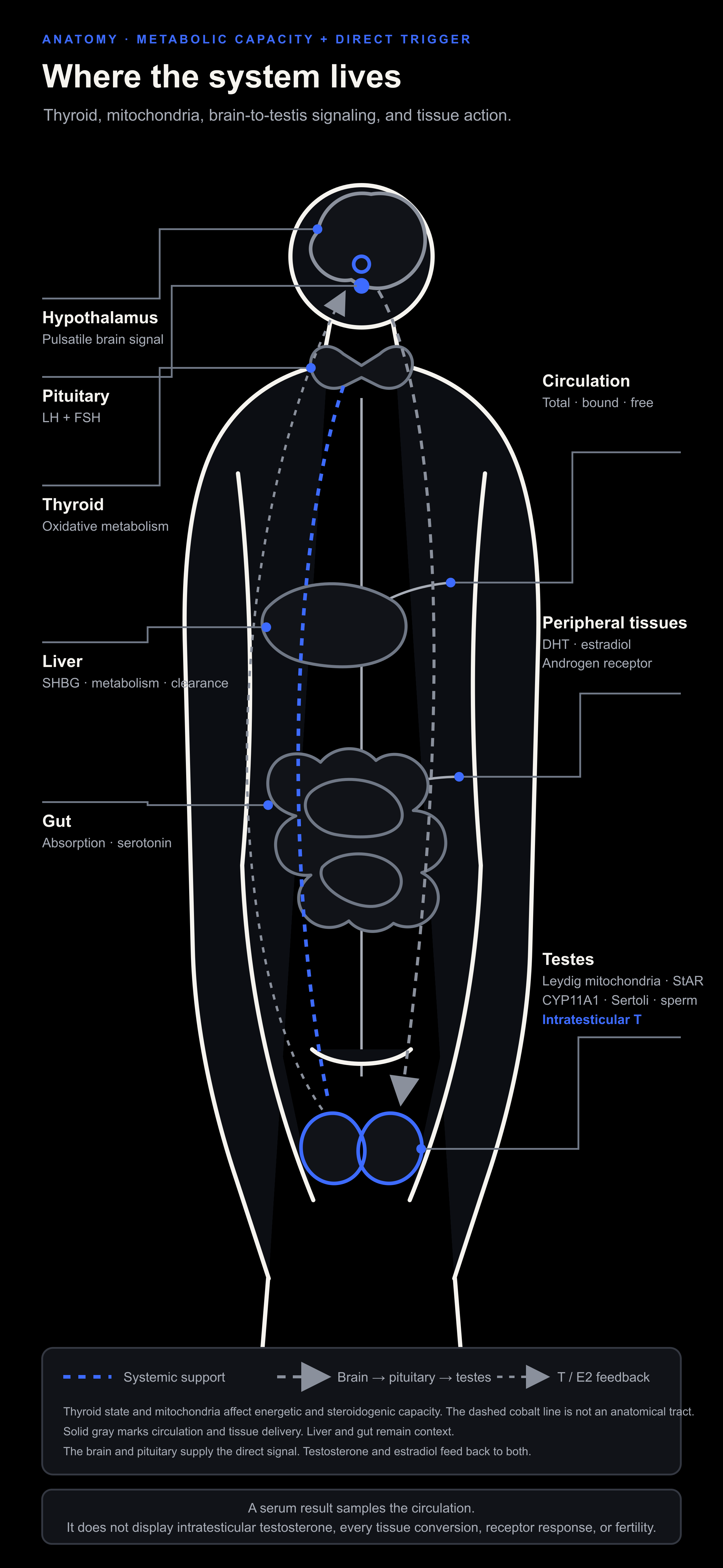
Figure 8. Dashed cobalt marks systemic metabolic support, not an anatomical tract. Gray shows the direct HPG signal, circulation, tissue delivery, and feedback. Liver and gut remain anatomical context.
Thyroid and mitochondria
Thyroid function changes the metabolic setting. Ray Peat argued that the pituitary is only one regulator of the thyroid system and that the liver, senses, nutrition, and stress also matter.[188] Thyroid disease can alter energy, SHBG, sex hormones, sexual function, and sperm parameters. Many of those changes improve when the thyroid disorder is treated.[189]
The useful human result comes from treating diagnosed disease. A before-after study followed forty treatment-naive men with overt primary hypothyroidism until they had remained euthyroid for six months on levothyroxine. Median testosterone rose from 162 to 441 ng/dL. Prolactin fell, sperm motility improved, and an assay-specific insulin-like factor 3, or INSL3, measure rose. The study had no untreated control, measured no free testosterone, and included only severe diagnosed hypothyroidism.[190] Its conclusion is specific: correct overt hypothyroidism. Euthyroid men have a different starting state.
Binding, transport, and peripheral conversion
Thyroid status is more than one TSH result. Thyroxine-binding globulin, or TBG, binds about three quarters of circulating thyroxine, or T4, and triiodothyronine, or T3. A change in TBG can move total thyroid-hormone values while free hormone and metabolic state remain stable.[191] This is the thyroid equivalent of reading total testosterone without its binding context.
The thyroid secretes mainly T4. About 80 percent of circulating T3 is produced outside the gland by conversion of T4.[192] D1 and D2 activate T4 to T3. D3 inactivates T4 and T3. Transporters move these hormones into cells, and local deiodinases can make tissue exposure differ from the serum result. Liver, kidney, brain, pituitary, muscle, illness, fasting, and medication can all change part of this handling.[192][193]
Read TSH with free T4, symptoms, illness, medication, and binding context. Use T3 or other tests when the clinical question justifies them. A total value changed by TBG is not a reason to force thyroid hormone, and a peripheral-conversion mechanism is not a general T3 protocol.
Thyroid hormone has no place as a general testosterone booster. Excess exposure can cause arrhythmia, bone loss, muscle wasting, anxiety, insomnia, and sexual dysfunction.[189]
Check the following:
Symptoms and signs compatible with thyroid disease.
TSH and free T4.
Medications, supplements, acute illness, or severe dieting that may alter the result.
Persistence of the testosterone pattern after diagnosed thyroid disease is treated.
Temperature and pulse add context, but neither can diagnose thyroid disease or determine a dose.
8. Steroid production
Cholesterol supplies the starting material. LH, mitochondrial transport, and enzymes determine what happens next.
Thyroid-supported metabolism and healthy mitochondria provide capacity. LH gives the Leydig cell its direct trigger. StAR moves cholesterol to the inner mitochondrial membrane, CYP11A1 converts it to pregnenolone, and later enzymes carry the pathway to testosterone.[85][86]
The brain-to-testis signal
The hypothalamic-pituitary-gonadal axis communicates in pulses. The hypothalamus releases gonadotropin-releasing hormone, or GnRH. The pituitary releases LH and FSH. LH gives Leydig cells the direct signal to make testosterone. FSH acts mainly on Sertoli cells and supports sperm production. Testosterone, estradiol, and inhibin B feed back to the brain and pituitary.[85][86][194]
The upstream pulse generator. Kisspeptin neurons sit above GnRH in the chain. In the arcuate nucleus, the KNDy network uses kisspeptin, neurokinin B, and dynorphin to help organize GnRH pulses. It integrates sex-steroid feedback with metabolic and environmental information.[195] In a small human infusion study, kisspeptin-10 increased LH and LH pulse frequency.[196] This maps the control system. Kisspeptin intervention belongs to research and specialist care.
The first diagnostic split. A low result may reflect a weak central signal or testes that cannot answer a strong one. LH and FSH help separate those patterns.[40][197]
The Kabbalah is wider than the HPG axis. The direct reproductive signal does not sit at the top of the whole map. Thyroid-supported metabolism, mitochondrial function, food, sleep, illness, and tissue integrity shape how well the testes answer it. Ray Peat described the pituitary as one part of a wider thyroid-regulating system.[188] His metabolic hierarchy surrounds the HPG signal without changing the physiology of LH.
Ray Peat’s hierarchy
“Many things, including the liver and the senses, regulate the function of the thyroid system.”[188]
Ray Peat’s official writing places thyroid, vitamin A, and low-density lipoprotein, or LDL cholesterol, among the conditions that support progesterone synthesis. It also places pregnenolone formation inside the mitochondrion.[198]
ATP supports cellular work. Reduced nicotinamide adenine dinucleotide phosphate, or NADPH, supplies the reducing power used in steroid synthesis. These are biochemical requirements, not blood targets or reasons to buy a generic mitochondrial stack.[85]
Pregnenolone sits upstream of dehydroepiandrosterone, or DHEA, progesterone, and the later steroids. The figure uses 17-OH as shorthand for 17-hydroxy.
Figure 9A preserves every item from the expanded source image. The blocker list is estrogen, non-esterified fatty acids, or NEFA, X-rays, serotonin, ultraviolet, darkness, PUFA, excess iron, prolactin, bacterial lipopolysaccharide, or LPS, carrageenan, and nitric oxide. The promoter list is thyroid, sugars, red light, vitamins A, D, E, and K2, glycine or gelatin, calcium, magnesium, caffeine, aspirin, stimulating life, some antibiotics, and saturated fats.
The source image also puts “EFA,” meaning essential fatty acids, beside PUFA.
The items in those lists carry very different levels of human evidence. Read the figure as Ray Peat’s metabolic model, not as a stack. Aspirin, thyroid hormone, antibiotics, and high-dose vitamins each require their own indication and safety review.
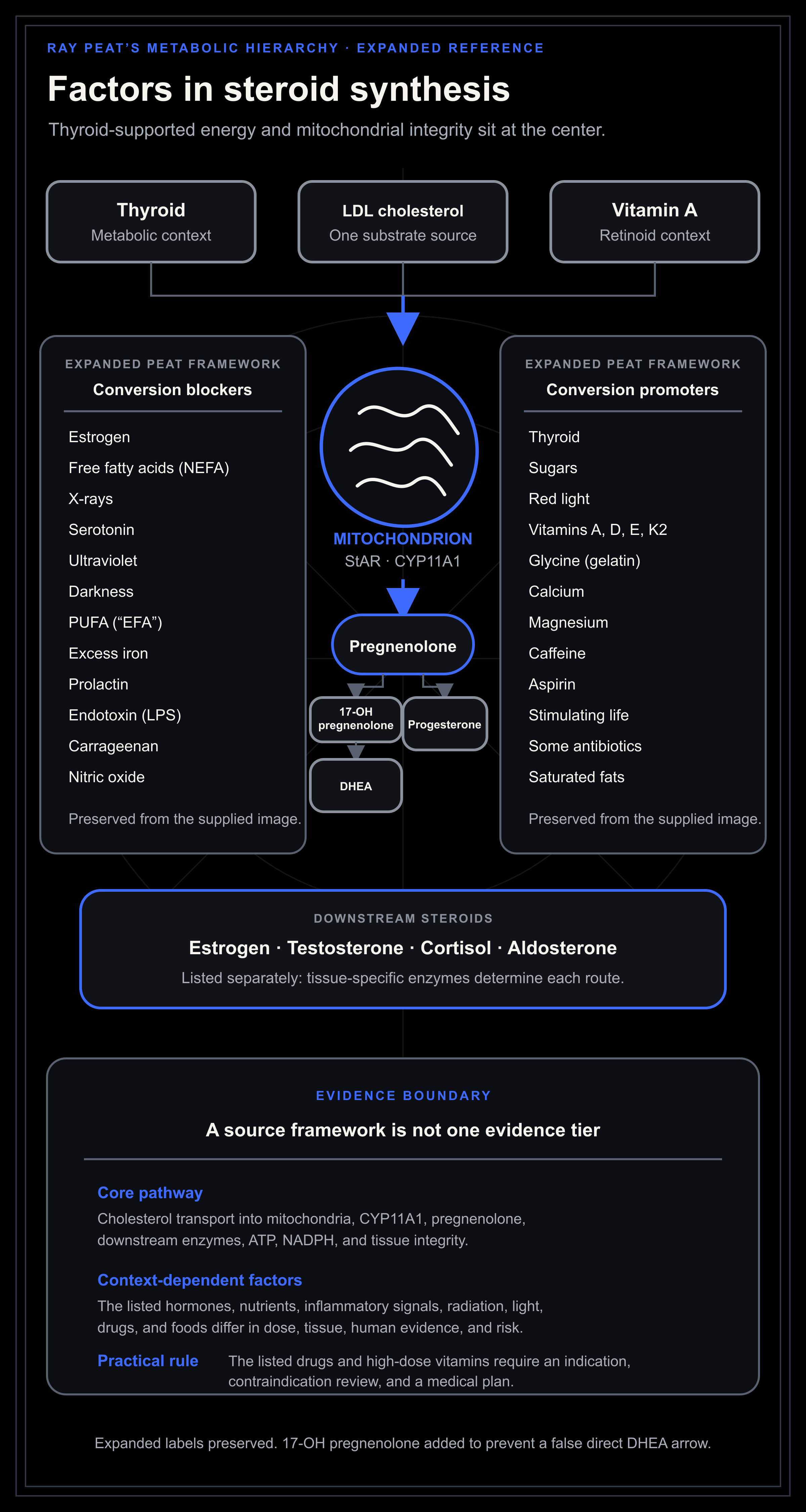
Figure 9A. Ray Peat’s metabolic hierarchy appears here with every item from the expanded reference. The source labels do not carry equal human evidence and should not be read as a supplement or drug protocol. The 17-OH pregnenolone intermediate is added so the DHEA branch remains biochemically correct.
The direct reproductive signal
GnRH arrives in pulses. The LH receptor activates cyclic adenosine monophosphate, or cAMP. cAMP activates protein kinase A, or PKA, which helps stimulate StAR-mediated cholesterol transport and steroid production.[85][86]
FSH acts mainly on Sertoli cells, where it supports sperm production and raises inhibin B.[194] Serum testosterone alone cannot answer a fertility question.
The cholesterol pool
Leydig cells can obtain cholesterol from several sources:
De novo synthesis from acetyl-CoA.
Uptake from circulating lipoproteins.
Stored cholesteryl esters in lipid droplets.
Mobilization from cellular membranes.
Carbohydrate, fat, and some amino acids can all supply acetyl-CoA, which the body can use to build cholesterol through the mevalonate pathway.[87][88] Cholesterol synthesis and testosterone production do not require a high-fat diet.
Dietary cholesterol supplies substrate while LH signaling, StAR transport, CYP11A1, later enzymes, and Leydig-cell health control its use.[85][86] Eat for nutritional adequacy. A dietary-cholesterol target cannot maximize the whole pathway.
The mitochondrial gate
Steroid production begins inside the mitochondrion by moving cholesterol to the inner membrane. Steroidogenic acute regulatory protein, StAR, controls much of this acute transport. Cholesterol side-chain cleavage enzyme, CYP11A1, then converts cholesterol to pregnenolone.[85][86]
A cholesterol-rich cell may still make little steroid because this step is regulated. Transport, enzyme expression, LH, mitochondrial integrity, and cofactors may each become the bottleneck.
Delta-5 and delta-4 routes
From pregnenolone, human steroid production follows related routes:
In the delta-5 route, pregnenolone becomes 17α-hydroxypregnenolone, then dehydroepiandrosterone, or DHEA. DHEA can reach testosterone through androstenediol or androstenedione.
In the delta-4 route, pregnenolone becomes progesterone, then 17α-hydroxyprogesterone, then androstenedione, then testosterone.
The main enzymes include cytochrome P450 17A1, or CYP17A1, 3β-hydroxysteroid dehydrogenase type 2, or HSD3B2, 17β-hydroxysteroid dehydrogenase type 3, or HSD17B3, and aldo-keto reductase, or AKR, family members. Their expression and cofactors determine the flow.[85]
ATP supports cellular work, while NADPH supplies reducing power for steroidogenic reactions.[85] A product labeled “mitochondrial support” neither identifies nor fixes the limiting step.
My first article also stressed glucose oxidation, carbon dioxide, redox balance, and restraint of excessive lipolysis.[80] The part worth keeping is that steroid production needs a working energy system. The ideal fuel ratio will differ between people.
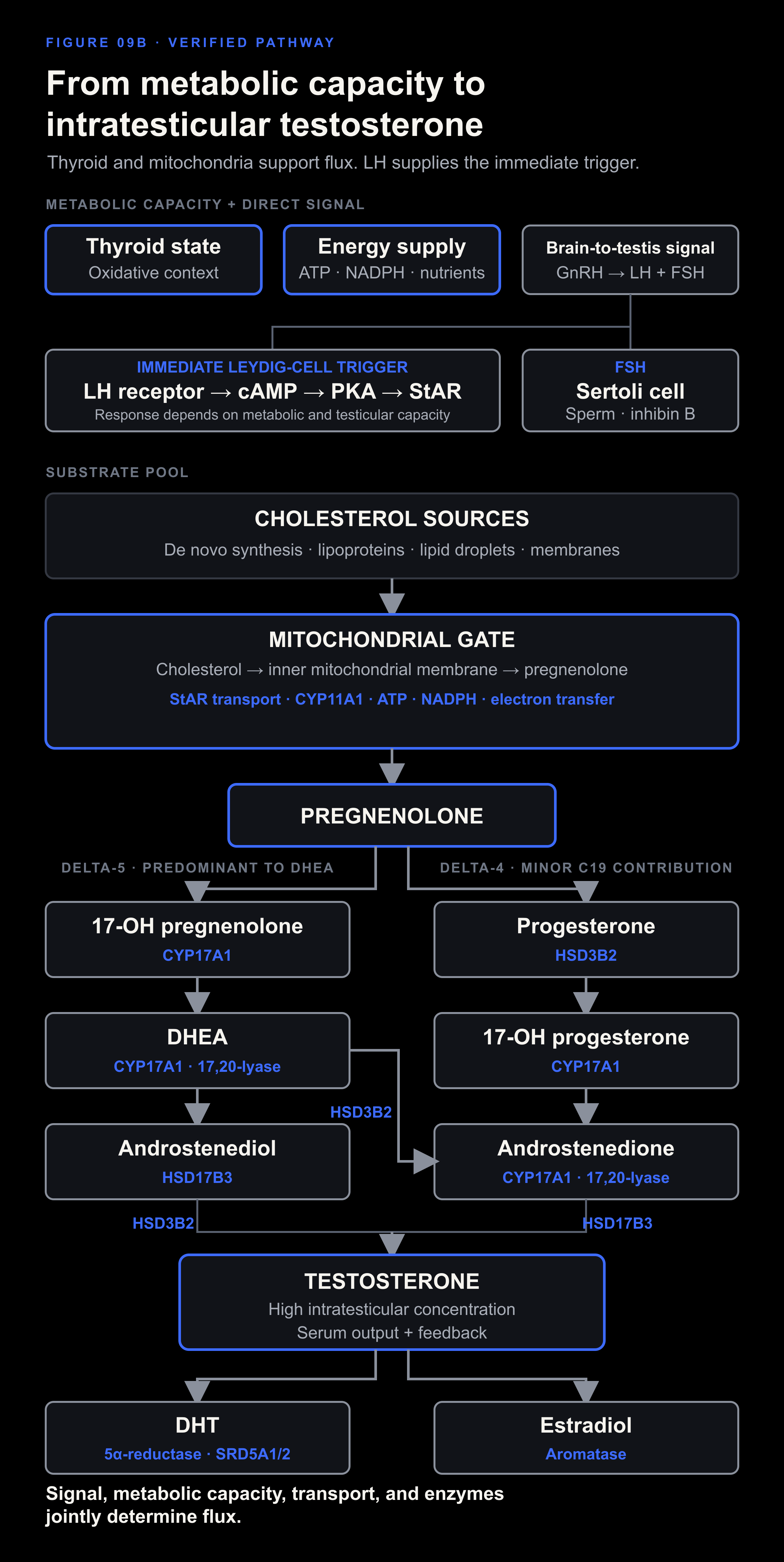
Figure 9B. Thyroid-supported metabolism and mitochondrial integrity set the metabolic capacity. LH supplies the immediate Leydig-cell trigger. StAR, CYP11A1, enzyme activity, reducing power, and testicular integrity determine the resulting flux.
Intratesticular testosterone
Testosterone reaches a much higher concentration inside the testis than in serum, and LH closely tracks that local steroid environment.[199] Spermatogenesis depends on the high intratesticular concentration.
In one controlled study of healthy men, exogenous testosterone lowered LH and FSH while intratesticular testosterone fell by about 94 percent.[200] Serum androgen exposure had risen as the concentration inside the testes collapsed.
That difference changes several decisions:
A serum testosterone result does not measure intratesticular testosterone.
Exogenous testosterone is not a fertility-preserving way to “restore natural production.”
A man who may want children needs to state that before starting androgen therapy.
Semen analysis answers a fertility question that serum testosterone cannot.
9. Liver, gut, serotonin, prolactin, illness, and stress
Liver, gut, immune, neurotransmitter, and stress pathways can change hormone transport, clearance, central signaling, testicular output, symptoms, and the meaning of a blood result.
Liver, SHBG, and clearance
The liver makes SHBG and metabolizes steroids. Liver disease, insulin resistance, thyroid status, and estrogen exposure can all change SHBG.[85][40]
On average, men with fatty liver have lower total testosterone and SHBG.[201] Adiposity and insulin resistance explain part of the association. Calling for a “liver detox” does not identify or treat the cause.
Review alcohol, medications, supplements, metabolic risk, liver enzymes, and imaging when indicated. Symptoms alone cannot measure estrogen clearance.
Gut and endotoxin
The gut influences nutrient absorption, bile acids, immune signaling, microbial metabolites, and illness burden.
A 2022 study identified a microbial enzyme capable of degrading testosterone.[202] The work combined isolates, animal experiments, and human associations. It establishes a mechanism worth following. Clinical use requires evidence of the organism, the enzyme, and a treatment that changes the outcome. Bloating alone supplies none of those.
Lipopolysaccharide can suppress Leydig-cell steroid production in animals.[203] A small study in overweight men also linked markers of endotoxemia with hypogonadism.[204] Together, these findings support treating diagnosed gut and metabolic disease, not rotating random antibiotics, antifungals, or probiotics.
For persistent gut symptoms, proceed conservatively:
Diagnose persistent symptoms such as bleeding, weight loss, anemia, chronic diarrhea, severe pain, or nocturnal symptoms.
Address celiac disease, inflammatory bowel disease, infection, constipation, reflux, or malabsorption when present.
Simplify obvious dietary triggers without collapsing nutritional adequacy.
Keep the diet nutritionally complete.
Reassess whether testosterone changed after the underlying disease improved.
Most serotonin is made in the gut
The intestine, mainly its enterochromaffin cells, produces about 95 percent of the body’s serotonin.[205] Peripheral serotonin remains separate from brain serotonin and helps regulate gut movement, secretion, sensation, platelets, and immune signaling.
Gut microbes can alter enterochromaffin serotonin production in animal and human microbial models.[206] Build the clinical picture from symptoms, disease, medication, diet, and physiology. A generic stool label of “high serotonin” does not represent the system.
The first article treated serotonin as a broad suppressive factor. The evidence supports a more specific account:
Gut serotonin can affect motility, secretion, pain, and inflammation.
Brain serotonin and peripheral serotonin are separated by the blood-brain barrier.
Receptor type and tissue decide the effect.
In cultured rat Leydig cells, serotonin activated a 5-HT2 and CRF route that reduced cAMP and testosterone production.[207]
Tissue, receptor, and compartment determine what a serotonin measurement means.
That experiment did not test gut serotonin, circulating serotonin, or living men. Review constipation, diarrhea, reflux, inflammatory disease, medication effects, and diet before attaching symptoms to serotonin. A generic anti-serotonin drug protocol has no place here.
Prolactin and sexual behavior
Persistently high prolactin can suppress GnRH, lower LH and testosterone, reduce libido, impair erections, and affect fertility.[67] Include it in the diagnostic workup when the pattern points there.
Record sodium adequacy in the same history. In Ray Peat’s model, low sodium can raise serotonin and sympathetic activity, serotonin can raise prolactin, and prolactin can promote further sodium loss.[90] Adequate salt can interrupt that indirect loop when sodium depletion is present. It cannot explain away a persistently high prolactin result or replace evaluation of medication, thyroid, kidney, or pituitary causes.
Orgasm causes an acute prolactin rise after masturbation and intercourse.[32][33] Repeated sessions repeatedly recreate the acute post-orgasm period. A properly timed blood test shows whether prolactin also remains elevated between sessions.
Intercourse and masturbation create different endocrine contexts. One paper pooled three earlier laboratory datasets. It compared separate groups rather than testing the same people twice. The total sample was 19 men and 19 women. Among the men, nine were studied after intercourse and ten after masturbation while watching an erotic film. Adjusted prolactin change was 15.62 ng/mL after intercourse and 3.05 ng/mL after masturbation. The authors interpreted the roughly fivefold difference as greater satiety.[208] This is a useful acute comparison. It does not establish a chronic hormone pattern or prove that modern internet pornography caused the difference.
Oxytocin also rises during self-stimulation and orgasm.[209][210] It adds another endocrine signal to the event. The available experiments do not show that oxytocin cancels prolactin or prevents receptor adaptation.
The receptor evidence comes from rats. In the main satiety model, sexually experienced males copulated for up to four hours. They reached at least five and an average of seven ejaculations before a 90-minute period without another ejaculation marked satiety. At 24 or 48 hours, 70 percent would not mate even with a novel female. Most copulated again by 72 hours, and the review describes full behavioral recovery by day 15.[34][35]
Androgen-receptor density changed by region. One ejaculation and full satiety both lowered it in the nucleus accumbens and medial preoptic nucleus at 24 hours. Only the fully satiated rats showed a reduction in the ventromedial nucleus, and the fully satiated group had lower density than the one-ejaculation group in the medial preoptic and ventromedial nuclei. Serum testosterone was similar across groups. A separate rat experiment found that ventral-prostate androgen receptors rose after the first three ejaculations and returned to baseline after the fourth.[211] Tissue, timing, and exposure change the response. Equivalent human brain and penile data are absent.
The same caution applies to 5-alpha-reduction. The human paper used several short drug experiments with only four to seven normal men per group. During an exogenous-testosterone experiment, metoclopramide raised prolactin and removed the normal relationship between the rise in testosterone and the rise in DHT. The authors interpreted this as lower peripheral testosterone-to-DHT conversion. In a separate group of 20 men receiving chronic neuroleptics, prolactin and testosterone were inversely correlated.[212] A rat experiment found greater LH-stimulated testicular 5-alpha-reductase activity during hyperprolactinemia.[213] These findings differ because tissue, duration, and treatment differ. Pornography and masturbation were not tested.
The receptor model comes from animal tissue because human brain and penile tissue have not been measured after porn or masturbation. Treat it as a mechanistic warning. The human case for removing porn stands independently on cue conditioning, compulsive repetition, sleep disruption, partnered-arousal mismatch, and sexual function.
Porn can train a narrow arousal environment. It can present a rapid stream of highly selected bodies, acts, angles, and scenes. With a wife or girlfriend, the body, pace, feedback, emotions, and uncertainty are real. They cannot be replaced with another cue at the movement of a finger.
Small experiments in men with compulsive sexual behavior found greater preference for novel sexual images and conditioned sexual cues.[214] Another small study found greater desire but similar liking for explicit videos, with stronger activity in a cue-reactivity network that included the ventral striatum.[215] These studies support a model of conditioned attention and wanting. They did not measure dopamine release.
That conditioning can create a mismatch. The screen produces an intense, controllable sequence of cues. Partnered sex asks the nervous system to respond to one real person. In a large cross-sectional survey of young men, higher problematic-pornography scores were associated with a higher probability of erectile dysfunction, while masturbation frequency was not a significant factor in the erectile-function model.[216] The cross-sectional design establishes association rather than direction. Across the broader review, problematic use carried more information than frequency alone.[36][37]
Remove porn during restoration. Stop edging. Give partnered arousal time to become responsive to partnered cues again. Bright screens near bedtime add a separate circadian and sleep load. The photons have the same effect whether the screen contains porn, work, or a film.
Run a defined restoration trial:
Remove pornography completely.
Stop edging and long compulsive sessions.
Pause or reduce masturbation when it leaves libido, erections, sleep, or focus worse.
Keep partnered sex separate in the record. It has a different sensory, relational, and satiety context.
Record morning erections, partnered arousal, mood, sleep, and recovered time.
Measure prolactin properly when symptoms or laboratory patterns point to it.
Changing the behavior may improve function even when prolactin is normal. A persistently high result still requires medical review. Medication, hypothyroidism, kidney disease, and pituitary disease are established causes.
Inflammation and illness
Acute illness can lower testosterone, while chronic inflammatory disease can affect the HPG axis, food intake, sleep, medication use, and testicular function.[40]
Routine diagnostic testing during severe acute illness can mislead. Treat the infection, inflammatory disease, organ dysfunction, medication effect, or energy deficit first.
Stress and glucocorticoid context
Glucocorticoids act at the hypothalamus, pituitary, and testes.[217] Sustained stress can also disturb sleep, appetite, training, substance use, and relationships.
One cortisol value cannot map the whole stress system. Sleep apnea, depression, overtraining, underfeeding, thyroid disease, Cushing syndrome, adrenal insufficiency, and medication effects each require their own diagnosis.
Include sodium intake and sodium loss in that map. Restriction can raise renin, aldosterone, and sympathetic catecholamines, while sodium intake also changes cortisol excretion and the meaning of a cortisol measurement.[89][91][218]
Start by removing loads that can be identified and measured:
Stabilize sleep and wake timing.
Restore adequate food if intake is low.
Reduce training when performance and recovery are falling.
Treat pain and illness.
Reduce alcohol and stimulant dependence.
Create periods without work demand.
Use psychological care when anxiety, trauma, depression, or compulsive behavior is the active constraint.
10. Conversion, transport, and tissue response
Testosterone is a branch point.
Progesterone and pregnenolone
Pregnenolone follows cholesterol directly. Progesterone sits in the delta-4 route to androgens and corticosteroids.[85]
My first article described progesterone as a counterweight to estrogen and stress. Supplemental progesterone adds a hormone to that model. Dose, route, conversion, feedback, and fertility determine whether the result helps or suppresses the male androgen state.
DHT
The main conversion. 5α-reductase uses NADPH to reduce testosterone to DHT. DHT binds the androgen receptor strongly and matters in external sexual development, prostate, skin, hair follicles, and other androgen-responsive tissues.[85][219]
The name 5α-reductase covers several isoenzymes. SRD5A1 has broad expression that includes liver and non-genital skin. SRD5A2 is prominent in androgen-sensitive reproductive tissues. SRD5A3 exists as a separate enzyme, although its normal contribution to adult androgen handling is less clear.[219] Finasteride inhibits type 2 more strongly. Dutasteride inhibits types 1 and 2. The drugs therefore do not create identical tissue effects.
Alternative routes. The shortest DHT route is testosterone to DHT. Other routes also exist. In the primary backdoor pathway, progesterone-related intermediates are reduced before CYP17A1 cleavage and proceed through androsterone and androstanediol to DHT. This route bypasses androstenedione and testosterone. A second route converts androstenedione to 5α-androstanedione and then to DHT, also bypassing testosterone.[85][220]
DHT is also a starting point for other metabolites. It can be reduced to 3α-androstanediol or 3β-androstanediol and can contribute to androsterone metabolites. Some reactions are reversible. Back-conversion of 3α-androstanediol to DHT has been shown in prostate tissue and studied heavily in prostate cancer.[220] That local adaptation should not be described as a universal systemic DHT recycling loop.
The liver, kidney, and target tissues then inactivate and conjugate androgens. UGT2B7, UGT2B15, and UGT2B17 help form glucuronides for urinary or biliary elimination.[219][221][222] These metabolites can reveal parts of androgen handling that a serum DHT value misses.
Blood and tissue are different compartments. In a randomized study of healthy men, a large increase in serum DHT left intraprostatic DHT and measured androgen-regulated prostate genes unchanged over four weeks.[223] Serum DHT describes blood exposure. Tissue response requires tissue-specific evidence.
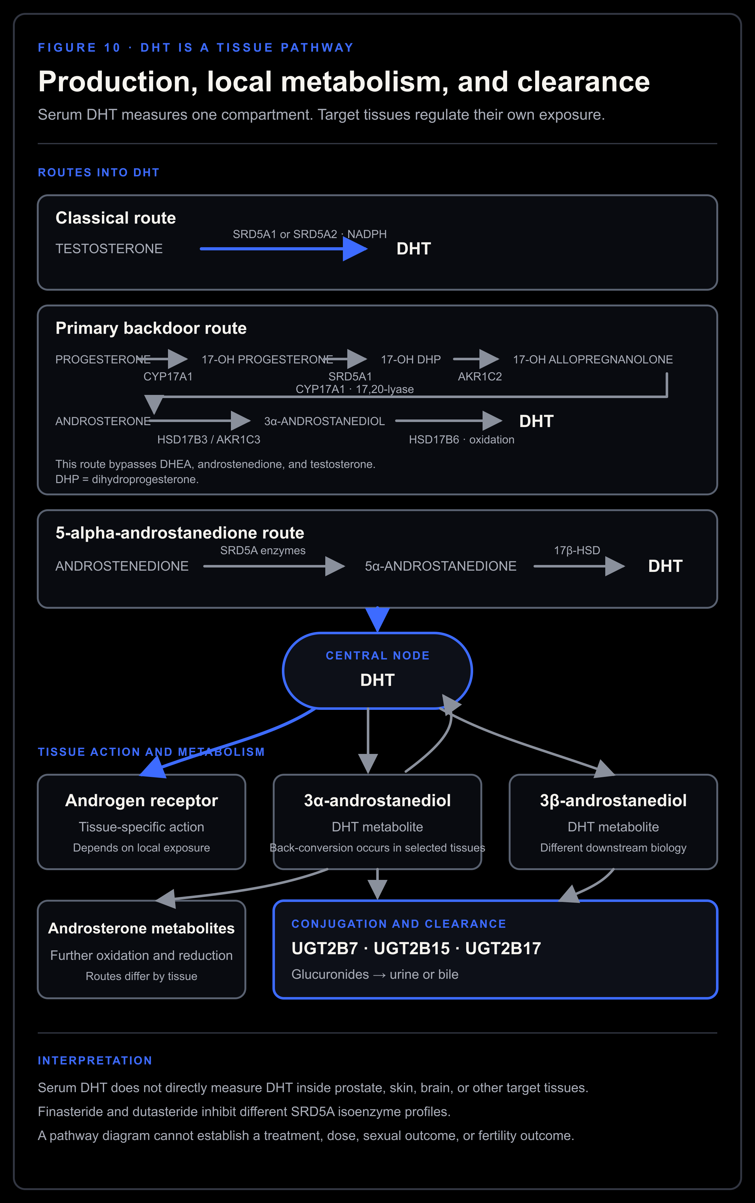
Figure 10. DHT can arise through testosterone reduction or routes that bypass testosterone. Local metabolites, back-conversion in selected tissues, conjugation, and clearance explain why serum DHT cannot stand in for every tissue.
The published pathway follows steroid-biochemistry reviews and human tissue studies. Claims about mood, fertility, pheromones, or a dosing protocol require their own evidence.
Because finasteride and dutasteride inhibit 5α-reductase, sexual symptoms, fertility plans, the original indication, and alternatives belong in the discussion with the prescriber. Do not stop a prescribed drug on the basis of one serum DHT result.
Estradiol
Aromatase converts testosterone to estradiol and is present in adipose tissue, which allows body composition to influence conversion. In men, estradiol supports bone, body composition, sexual function, and hormonal feedback.[85][27]
In a controlled experiment, androgen deficiency drove losses in lean mass, muscle size, and strength, while estrogen deficiency contributed to fat gain. Both deficiencies affected sexual function.[27]
The sexual result changes when a strong nonaromatizable androgen replaces testosterone. In a two-year randomized trial, pharmacological DHT suppressed testosterone, LH, FSH, and estradiol while leaving most self-reported sexual measures unchanged. Desire fell slightly, and spinal bone density declined.[224] High DHT exposure can preserve part of the sexual signal while losing estrogen-dependent bone protection. Indiscriminate aromatase suppression follows the same biological mistake.
Very high estradiol can matter in the right context. Driving it as low as possible can impair bone, lipids, sexual function, and body composition.
SHBG and free testosterone
Most circulating testosterone travels bound to SHBG or albumin, leaving a smaller fraction free.[40]
Some direct free-testosterone tests are unreliable. Equilibrium dialysis serves as a reference method, while validated calculations can help when total testosterone, SHBG, and albumin are measured accurately.[40]
Low SHBG often accompanies obesity, insulin resistance, and hypothyroidism. Hyperthyroidism, liver disease, some medications, and estrogen exposure may raise it. SHBG helps interpret the result without becoming the center of the Kabbalah.
Receptor response, symptoms, and function
Androgen action depends on receptor function, tissue exposure, local metabolism, illness, training, nutrition, and the many non-androgen causes of the same symptoms.
Routine clinical practice has no test for everyday “androgen receptor sensitivity.” Supplement claims about it usually rest on indirect or animal data.
Use symptoms and function carefully:
Libido is influenced by androgens, estradiol, dopamine, mood, relationship context, medications, sleep, and illness.
Erections require vascular inflow, smooth-muscle relaxation, intact nerves, adequate sleep, and psychological context.
Muscle gain requires training stimulus, food, amino acids, recovery, and time.
Fertility requires gonadotropins, intratesticular testosterone, Sertoli-cell function, sperm production, and unobstructed transport.
Energy and mood are broad outputs with many non-androgen causes.
Fatigue and mood have many causes. Libido, morning erections, gonadotropins, fertility, and matched laboratory values offer a sharper androgen picture.
Part II. Read the laboratory pattern
11. Laboratory interpretation
Hypogonadism requires compatible symptoms or signs together with consistently low testosterone. The diagnostic standard uses accurate assays and a repeated morning fasting total testosterone measurement. Free testosterone becomes useful when total testosterone sits near the lower limit or SHBG is altered.[40]
Do not diagnose hypogonadism from one result.
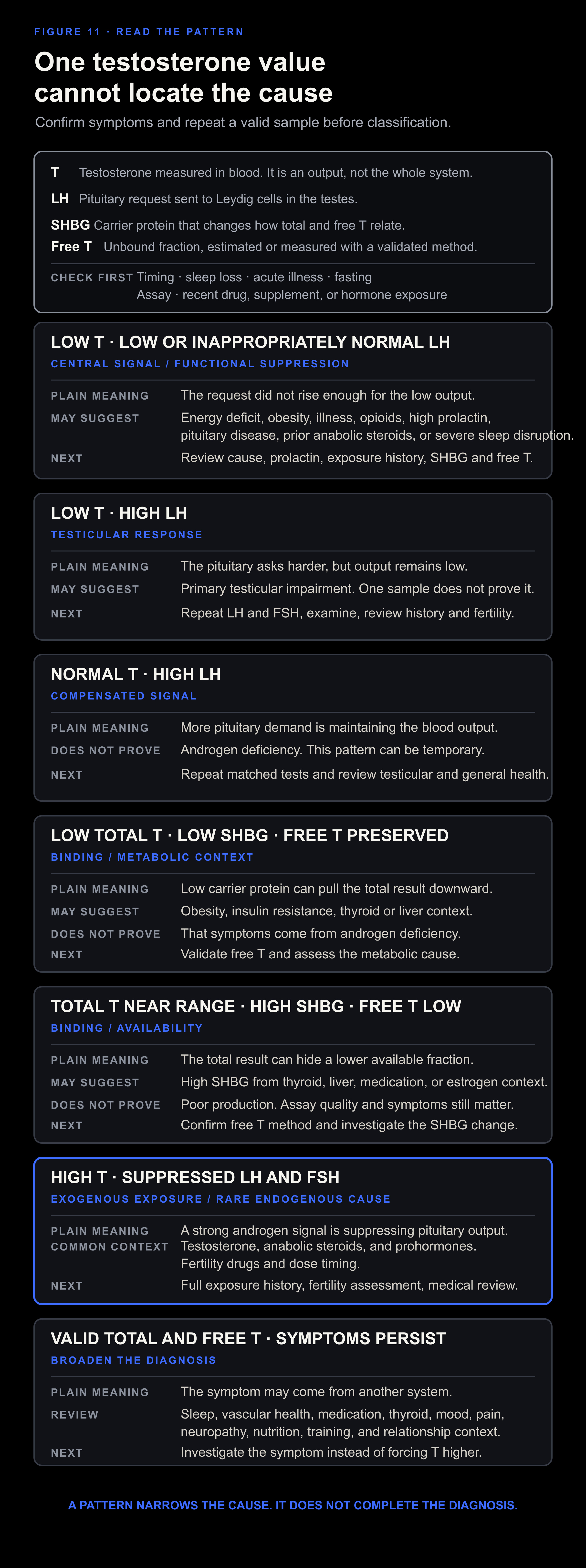
Figure 11. Pair testosterone with symptoms, timing, SHBG or free testosterone, and gonadotropins.
A valid baseline
Repeat a first low result under ordinary, comparable conditions:
Draw after a representative sleep period.
Use a morning sample for men on a conventional schedule.
For shift workers, coordinate interpretation with sleep timing and clinician judgment.
Use the same reliable laboratory when possible.
Fast when following the diagnostic standard.
Avoid testing during acute illness.
Record medications, supplements, androgen exposure, and recent injections or gels.
Avoid drawing immediately after an unusual all-night event, extreme endurance session, or severe caloric depletion.
Use the reference range for that assay and laboratory.
The core laboratory set
The core workup for repeated low testosterone often includes:[40][197][67]
Total testosterone.
SHBG.
Albumin for calculated free testosterone, or a validated free-testosterone method.
LH.
FSH.
Prolactin.
Add tests according to the likely cause:
TSH and free T4 when thyroid disease is possible.
Complete blood count.
Comprehensive metabolic panel and liver enzymes.
Ferritin and transferrin saturation when iron deficiency or iron overload is possible.
Hemoglobin A1c or fasting glucose for metabolic risk.
Semen analysis when fertility is the question.
INSL3 when Leydig-cell capacity remains uncertain and the assay is available.
Pituitary hormones or imaging when secondary hypogonadism, hyperprolactinemia, headache, visual symptoms, or other pituitary deficits are present.
Testicular ultrasound when examination identifies a mass or structural concern.
Read LH beside testosterone
LH reflects demand. Testosterone reflects one part of output. Reading them together shows how much signal accompanies the measured result.
The favorable pattern is adequate testosterone and testicular function with less compensatory LH demand. Low LH by itself is not the goal. Low LH with low testosterone points toward a central or functional problem. Stable testosterone that requires progressively more LH points toward reduced testicular response.
A lower LH-to-testosterone ratio means that less measured LH accompanies the same testosterone. A higher testosterone-to-LH ratio expresses the same relationship in the opposite direction. Name the direction and the units because the number changes when testosterone is reported in ng/dL rather than nmol/L.
Among 8,182 young European men with a median age of 19.1 years, testosterone alone did not differ across sperm-concentration groups. Higher LH accompanied lower sperm concentration, total sperm count, motility, and percentage of normally shaped sperm. Both total-testosterone-to-LH and calculated-free-testosterone-to-LH ratios rose with the same semen measures. The pattern extended into semen values that the authors considered normal, and the LH difference became clear below roughly 70 to 80 million sperm per milliliter compared with higher categories.[225]
This was a cross-sectional association. It does not prove that lowering LH improves fertility, and it does not create a universal ratio target. It shows why a normal testosterone result can hide greater pituitary demand.
Trend the relationship. The ratio becomes more useful across repeated tests performed under similar conditions. Stable natural testosterone with rising LH suggests that the testes require more pituitary demand. Stable testosterone with repeatedly lower LH shows the same output alongside less measured demand. An LH pulse, sampling time, illness, sleep loss, or an assay change can distort any single ratio.
INSL3 and Leydig-cell capacity
INSL3 reflects Leydig-cell capacity more than immediate output. Mature Leydig cells secrete it, and its level varies less within one person than testosterone.[226][227]
A blood test measures circulating INSL3. The term INSL3 expression belongs to gene or tissue measurements.
INSL3 can add context after anabolic-steroid exposure. Former users had lower levels than controls years after cessation, even after accounting for total testosterone.[228] Testosterone may therefore recover while Leydig-cell capacity remains reduced.
Keep the proposed INSL3 cutoff inside its population and assay. A 2024 analysis proposed 0.4 ng/mL or lower as a low value in middle-aged and older men. It corresponded to the lower 95 percent boundary around age 60 to 65 in the analyzed populations. After men at or below that threshold were removed, the authors proposed a serum reference interval of 0.4 to 2.3 ng/mL for men older than 35.[227]
The number is not universal. INSL3 fell by about 14 percent per decade in the EMAS data. Plasma averaged about 10 percent below serum. Different assays produced materially different ranges. At the 0.4 ng/mL threshold, the comparison with low overall sexual function still produced 41.0 percent false positives and 24.1 percent false negatives. The paper therefore supports INSL3 as context for Leydig-cell capacity, especially when testosterone and LH tell an incomplete story. It does not support using one INSL3 result as a diagnosis.[227]
Assay access remains limited, and INSL3 belongs after the standard laboratory set. When available, read it beside testosterone, LH, testicular volume, semen analysis, specimen type, age, and the clinical question.
Pattern 1: low testosterone with low or inappropriately normal LH
Low testosterone with low or inappropriately normal LH points toward a secondary, central, or functional pattern. The pituitary signal has not risen appropriately for the low output.
Possible contexts include obesity, energy deficiency, acute or chronic illness, opioid use, hyperprolactinemia, pituitary disease, prior anabolic-steroid use, glucocorticoid exposure, severe sleep disruption, and some genetic conditions.[40][197][229][230]
The word “normal” can mislead. An LH value inside the laboratory range may be physiologically inappropriate when testosterone is low.
Pattern 2: low testosterone with high LH
Low testosterone with high LH points toward primary testicular impairment. The pituitary has increased its signal, but the testes have not produced an adequate output.
Possible causes include genetic conditions, prior orchitis, torsion, trauma, chemotherapy, radiation, advanced testicular damage, or loss of testicular tissue. FSH, examination, fertility history, and semen analysis help define the problem.[40][197]
Pattern 3: low total testosterone, low SHBG, and preserved free testosterone
Obesity and insulin resistance can produce this combination. Sometimes the result reflects a binding-protein pattern more than severe androgen deficiency. Symptoms, repeated measurements, assay quality, and metabolic context determine its significance.
The pattern gives a reason to improve metabolic health while continuing to investigate symptoms. It gives no reason to pursue the lowest possible SHBG.
Pattern 4: total testosterone near range, high SHBG, and low free testosterone
High SHBG can keep total testosterone near the laboratory range while the available fraction is low. Hyperthyroidism, liver disease, estrogen exposure, some medications, low body weight, and age can contribute to high SHBG.[40]
Confirm the free-testosterone method, then investigate the SHBG change. This is a binding pattern first. Testicular production remains a separate question.
Pattern 5: normal testosterone with high LH
This pattern has been called compensated hypogonadism. Longitudinal data show frequent reversion, so the finding marks increased pituitary demand rather than a settled diagnosis of symptomatic androgen deficiency.[197][231]
The pattern can signal reduced testicular reserve, broader health deterioration, or differences in hormone handling. In a cross-sectional study of 1,865 men, aromatase haplotypes were associated with estradiol and LH in opposite directions. The LH differences were strongest in men aged 65 or older.[232] High LH still records increased pituitary demand. The ratio gains meaning only beside symptoms, repeat measurements, fertility data, and testicular context.
Repeat testing and a review of the cause are more useful than immediate hormone escalation.
Pattern 6: high testosterone with suppressed LH and FSH
Exogenous androgen exposure commonly explains high testosterone with suppressed LH and FSH. Interpretation depends on timing relative to an injection, gel, oral product, or anabolic agent. Rare endogenous causes still require medical evaluation.
Ask directly about prescribed testosterone, underground products, prohormones, selective androgen-receptor modulators, shortened to SARMs, human chorionic gonadotropin, or hCG, fertility drugs, and contaminated supplements. Full disclosure prevents misclassification.
Pattern 7: valid total and free testosterone with persistent symptoms
When repeated measurements are appropriate and the androgen pattern remains normal, widen the diagnosis. Erectile symptoms may be vascular, neurological, medication-related, sleep-related, or psychological. Fatigue and low mood may come from thyroid disease, anemia, pain, depression, sleep disruption, inadequate food, or another illness.
The next test should follow the symptom. Forcing testosterone above an adequate physiological result does not identify the cause.
Prolactin
High prolactin can suppress GnRH and gonadal function. Pituitary adenoma, medication, hypothyroidism, kidney disease, and physiological stress all belong among the possible causes.[67]
A mild elevation often needs an unhurried repeat. When possible, avoid drawing immediately after sex, hard exercise, or a stressful venipuncture, and review medication and thyroid status.
Persistent elevation requires evaluation. Headache, visual change, galactorrhea, or other pituitary deficits make that evaluation more urgent. Dopamine agonists require a defined cause and medical management.
Measure prolactin and androgens when the pattern calls for them. Remove pornography during the restoration phase regardless of the prolactin result. Stop edging and review compulsive masturbation because the behavior can damage sexual function, sleep, and attention even when basal prolactin is normal.
Four patterns in real life
The underfed athlete. A man loses weight quickly, adds intervals, sleeps worse, loses libido, and gets a low testosterone result with low or inappropriately normal LH. The pattern points toward reduced central drive in an energy and recovery deficit. The first action is to restore energy availability and reduce load. A booster that pushes LH would leave the cause in place.
The man with obesity and sleep apnea. He has low total testosterone, low SHBG, preserved calculated free testosterone, loud snoring, and daytime sleepiness. The blood result partly reflects low binding protein. Adiposity and fragmented sleep remain the stronger upstream targets. He still needs a proper clinical interpretation because symptoms and free testosterone decide how much of the pattern is androgen-related.
The man with a testicular pattern. He has repeated low testosterone with high LH, a history of torsion, and a fertility goal. The pituitary is already sending a strong request. More LH-oriented stimulation cannot rebuild lost testicular tissue. Examination, follicle-stimulating hormone, semen analysis, and reproductive care take priority.
The man with normal androgen measurements and persistent symptoms. He has two valid testosterone results, appropriate free testosterone, and no abnormal gonadotropin pattern, yet erections and energy remain poor. The diagnostic branch widens to vascular health, medication, neuropathy, depression, sleep, pain, thyroid disease, relationship context, and other causes. Raising an already adequate testosterone result would test the wrong system.
12. Causes, exposures, and age
Foundational work must never delay evaluation of structural, genetic, medication-related, or serious disease.
Testicular causes
The relevant history includes undescended testes, delayed puberty, infertility, mumps orchitis, torsion, trauma, infection, testicular cancer, chemotherapy, pelvic or testicular radiation, and surgery. Examination may reveal small testes, asymmetry, varicocele, or a mass.[40]
Acute severe testicular pain is an emergency because torsion is time-sensitive. A painless mass also warrants prompt assessment.
Pituitary and hypothalamic causes
Secondary hypogonadism can arise from pituitary tumors, hyperprolactinemia, infiltrative disease, head trauma, cranial irradiation, genetic conditions, hemochromatosis, severe systemic disease, or medications.
A new severe headache, visual-field change, cranial nerve symptoms, vomiting with neurological change, or multiple pituitary deficits all require urgent attention. Lifestyle work comes later.
Medication and substance causes
Opioids are a major cause of gonadal suppression. A systematic review and meta-analysis estimated a high prevalence of hypogonadism among male users, although definitions and study results varied widely.[229] Management begins with medication review, coordination of pain care, and appropriate endocrine testing. Abrupt discontinuation may be unsafe.
Heavy or chronic alcohol exposure can suppress testosterone and damage sperm production through testicular, central, liver, sleep, and nutritional pathways.[233] Human findings at low or moderate intake are less consistent. Judge the exposure by its effect on sleep, liver function, nutrition, sexual function, and the hormone pattern rather than assuming one harmless dose fits every man.
Glucocorticoids, some antipsychotics, medications that raise prolactin, and other drugs can contribute. Finasteride, dutasteride, antidepressants, antihypertensives, alcohol, cannabis, and nicotine can affect sexual symptoms through pathways that may or may not involve serum testosterone.[40][67][217]
A complete medication list includes prescriptions, nonprescription drugs, supplements, injections, topical products used by a partner, and substances obtained outside medical care.
Environmental and occupational exposures
Environmental chemicals can interact with steroid synthesis, receptors, binding proteins, metabolism, and reproductive development. Human evidence varies widely with the chemical, dose, life stage, and outcome.
Recent meta-analyses associate higher exposure markers for some phthalates, bisphenol A, and per- and polyfluoroalkyl substances, or PFAS, with changes in testosterone, SHBG, or sperm outcomes.[234][235][236][237] Most contributing studies are observational. Urinary metabolites may represent a short exposure window, whereas serum concentrations of other chemicals can reflect long-term accumulation. Diet, occupation, adiposity, age, fertility status, and socioeconomic conditions can confound the results. Even compounds from the same chemical family may move an outcome in different directions.
The evidence supports targeted reduction of named exposures. Diagnose the chemical, source, and biological pattern before attributing symptoms to endocrine disruption. Commercial detox language skips all three.
Start with exposures that can be named and controlled:
Identify occupational exposure to pesticides, solvents, plasticizers, metals, heat, radiation, or combustion products.
Follow workplace controls, ventilation, protective equipment, and exposure monitoring.
Avoid smoking and unnecessary combustion exposure.
Store and heat food in materials designed for the temperature and use.
Replace damaged food-contact containers and avoid an impossible pursuit of complete plastic elimination.
Follow local fish, water, and contamination advisories when a specific exposure is known.
Discuss biomonitoring with occupational or environmental medicine when the exposure is substantial and the result would change management.
Use low-cost household changes that reduce a named exposure. Treat their testosterone effect as secondary and keep major findings in priority order: high LH, a pituitary pattern, severe OSA, opioid exposure, and sustained underfueling come first.
Anabolic-androgenic steroids and testosterone
Anabolic-androgenic steroids, shortened to AAS, suppress LH and FSH. A systematic review found substantial and sometimes prolonged effects on testosterone, gonadotropins, sperm production, and fertility.[230] The pooled hormone comparisons were small, with 17 to 65 users depending on the outcome. Exposure, dose, assay, and follow-up varied, and most studies had an unclear risk of bias. The suppression is clear. The recovery clock remains specific to the exposure and the man.
Some former users continue to have lower testosterone and symptoms years after stopping.[238] A second systematic review found 168 cases that it attributed to AAS-induced hypogonadism. Complete outcomes were known for only 38, and four met its definition of full HPG-axis recovery, including two after drug treatment.[239] That is not a population recovery rate. The evidence set was dominated by selected cases, missing outcomes, mixed exposures, and likely publication bias.
The prospective HAARLEM study followed one hundred men before, during, and after a self-directed androgen cycle. Testosterone returned to the laboratory range within three months after cessation in most men, while sperm recovery took much longer. At the final follow-up, 11 percent still had low testosterone and 34 percent still had a total sperm count below 40 million.[240] The last visit occurred about one year after the cycle began, not one year after it ended. Most men used several underground products, and post-cycle treatment was not randomized.
Recovery depends on the compounds, dose, duration, age, baseline function, and time since cessation. The reviews and the prospective cohort show wide variation. An internet post-cycle template ignores the variables that determine recovery. hCG, selective estrogen-receptor modulators, aromatase inhibitors, and testosterone each alter the axis differently and require medical supervision.
Age
Do not assign a low testosterone result to age until reversible causes have been checked.
Over time, men often accumulate poor sleep, excess fat, diabetes, inactivity, medication exposure, illness, undernutrition, and testicular damage. Each can lower testosterone. In a Baltimore longitudinal cohort, age was no longer significant after the model included several health conditions.[241] A clinical review likewise found little decline before advanced age among men without obesity, adverse lifestyle, or opioid use.[242]
Some longitudinal studies find an additional independent decline, especially in bioavailable testosterone and at advanced age. That effect can coexist with reversible causes. Check the reversible causes first.
Classify the pattern at any age. Check sleep, body composition, disease, medication, LH, FSH, SHBG, prolactin, thyroid status, and testicular history.
Part III. Restore the system
13. The natural restoration plan
Follow the steps in order. Nothing later in the protocol can repair an invalid first measurement or compensate for an untreated structural cause.
Gate 1: Validate and classify
Begin by proving that the symptom and laboratory pattern are real. Record the change from baseline, its onset, the fertility goal, and every relevant exposure. Repeat a low testosterone result under comparable conditions. Add SHBG or a valid free-testosterone measure when needed, then read LH, FSH, and prolactin beside it.[40][197][67][225][227]
An unreliable assay, unmatched repeat, or incomplete exposure history leaves the pattern unclassified. Red flags, structural clues, and fertility threats go directly to clinical care.
Step 1. Complete the symptom and cause intake
Complete the opening intake, including the change from baseline, its onset, and what happened before it.[40][41]
Step 2. Standardize the first measurement
Use an accurate laboratory and test after representative sleep. For a conventional schedule, draw in the morning while fasting and outside acute illness. Record wake time, diet phase, training, medication, and androgen exposure.[40][50]
Step 3. Confirm persistence
Repeat any low result under comparable conditions, adding SHBG and valid free testosterone when needed.[40]
Step 4. Classify the layer
Add LH, FSH, and prolactin, then classify the pattern. When fertility matters, include reproductive history and semen analysis early.[197][67][225][227]
Gate result: A repeated, usable testosterone result has been classified as central, testicular, binding-related, prolactin-related, exogenous, or still unresolved. Red flags move directly to care.
Gate 2: Stabilize circadian rhythm and sleep
Once the baseline is usable, stabilize sleep and circadian timing. Track wake-time variation, sleep opportunity, awakenings, daytime sleepiness, snoring, nasal obstruction, light timing, and the main disruptor.[46][42][25][56][64][62]
The direction should be clear: steadier timing, fewer avoidable awakenings, better mornings, and less daytime sleepiness. Persistent insomnia, suspected apnea, unsafe sleepiness, or structural nasal obstruction needs its own clinical branch.
Step 5. Use a stable wake time
Choose a workable wake time and keep it stable when possible. Shift workers should protect complete sleep cycles within the most stable schedule their rotation permits.[44][50]
Step 6. Build day-night light contrast
Let outdoor light reach the eyes after waking when feasible. Keep the day bright, reduce blue-rich light near sleep, and keep the bedroom dark. Shift work and circadian disorders may require different timing.[42][43][64][54]
Step 7. Protect complete sleep cycles
Reserve enough time for complete cycles, protecting the slow-wave-rich early sleep period and the REM-rich later sleep period. Persistent insomnia deserves evidence-based care.[46][49][48][45][47]
Step 8. Screen sleep disruptors
Review snoring, apnea signs, nasal obstruction, allergies, pain, reflux, restless legs, caffeine, alcohol, nicotine, heat, and noise.[25][56][58][57]
Safe saline rinsing and nasal strips can help when they fit the nasal problem. Mouth taping has no place with obstruction or suspected apnea.
Gate result: Sleep has enough opportunity, a usable schedule, and a clear day-night light pattern. Persistent insomnia, apnea signs, or nasal obstruction has entered a specific diagnostic branch.[62][61][59]
Gate 3: Restore energetic and metabolic conditions
Next, match food and body composition to the starting problem. Track weight, waist, appetite, performance, recovery, sleep, libido, and the relevant laboratory values. The aim may be refeeding, gradual fat loss, or correction of a confirmed disease or deficiency.[74][83][84][1][77][242]
Continued involuntary loss, crash dieting, worsening recovery, uncontrolled gain, or no response means the plan is wrong or incomplete. Eating-disorder signs, malabsorption, anemia, iron overload, diabetes, thyroid disease, liver disease, and persistent abnormalities require the relevant clinician or dietitian.
Step 9. Reconstruct the energy history
Reconstruct the previous twelve months of weight, waist, dieting, training, illness, appetite, and food restriction. A low result after a prolonged cut starts with a different likely explanation from one that appeared during weight gain with severe insulin resistance.[74][83]
Step 10. Correct the energy state
When low energy availability is likely, increase food and reduce unnecessary expenditure. Build the diet from complete protein, tolerated carbohydrate, enough fat, and varied micronutrient sources.[74][76][77]
The body can make cholesterol from carbon supplied by carbohydrate, fat, and amino acids, so high fat is unnecessary for steroid substrate.[87][88]
Step 11. Reduce excess adiposity without creating a crash diet
When obesity and insulin resistance dominate the pattern, combine a sustainable energy deficit with resistance training, walking or aerobic work, adequate protein, and clinical obesity care when appropriate.[83][84]
Aim for better metabolic health without sacrificing recovery.
Step 12. Review minerals and correct defined needs
Investigate anemia, iron overload, vitamin D deficiency, diabetes, thyroid disease, liver disease, kidney disease, and malabsorption. Then review calcium, magnesium, potassium, sodium, iodine, selenium, zinc, copper, vitamins A, D, E, and K, and the B vitamins.[21][189][1][190][102]
For sodium, review sweat, heat, endurance work, diarrhea, food restriction, blood pressure, kidney and heart function, and medication. For thiamine, review food intake, alcohol, malabsorption, prolonged vomiting, high carbohydrate demand, diuretic use, neuropathic symptoms, and persistent constipation. Correct the identifiable constraint. Keep gram-dose thiamine and aggressive salt loading outside the default protocol.[90][92][93]
Use the supplement map in Section 5. Match magnesium form to elemental dose and bowel tolerance. Use iodine and selenium only after the thyroid and intake context are clear. Read zinc, P5P, and vitamin E as conditional prolactin tools rather than a universal anti-prolactin stack. Keep glycine and L-theanine in the sleep-support category. Keep TMG, TUDCA, D-chiro-inositol, methylene blue, Epithalon, Pinealon, Vilon, and disease-level doses outside the default stack.[103][108][97][152][158][162][117][122][126][116][98][163][161]
Reserve supplements for a defined need. Change one main input at a time and define the stop rule before starting. Orange juice, raw carrot salad, well-cooked white button mushrooms, or well-boiled bamboo shoots can be tested consistently when they fit the energy, estrogen, and bowel pattern.
Gate result: Food intake and weight change now fit the starting problem. Weight, waist, performance, sleep, and symptoms show whether refeeding, gradual fat loss, or correction of a defined deficiency is working.
Gate 4: Adjust training and remove suppressors
After food and sleep can support adaptation, make training recoverable and complete the exposure history. Track strength, work capacity, soreness, tissue tolerance, sleep, libido, medication, alcohol, nicotine, hormones, compulsive behavior, and active illness.[30][31][67][229][240][233]
Performance and recovery should improve as each suppressor receives a clear plan. Dependence, psychiatric risk, medication withdrawal, organ disease, bleeding, severe pain, and fertility risk require supervised care.
Step 13. Program training for adaptation
Use progressive resistance training with enough aerobic activity to improve fitness. Hard sessions must remain recoverable. Transient post-workout testosterone is not a useful program metric.[30][31][187][185]
Step 14. Match volume to recovery
When performance, mood, libido, sleep, and tissue tolerance decline together, reduce volume or intensity and reassess food and illness. If training load is the suspected stressor, a deload also serves as a diagnostic intervention.[186]
Step 15. Review drugs, hormones, and compulsive habits
Make a complete list of opioids, glucocorticoids, antipsychotics, antidepressants, finasteride or dutasteride, alcohol, cannabis, nicotine, testosterone, anabolic steroids, SARMs, prohormones, hCG, fertility drugs, and supplements.[67][229][230][233]
Treat an unexpectedly strong bodybuilding or sexual-enhancement supplement as a possible exposure to an undeclared drug until the product is verified.[38][39]
Remove pornography during the restoration phase. Stop edging and long compulsive sessions. Pause or reduce masturbation when it impairs sleep, attention, libido, erections, or partnered arousal. Keep partnered sex separate in the record because its physiological and relational context differs.[36][37][208][209]
Discuss medication changes with the relevant prescriber. Dependence-forming and psychiatric drugs should never be stopped abruptly without appropriate care.
Step 16. Treat active illness and gut disease
Investigate persistent pain, gastrointestinal disease, autoimmune disease, infection, dental disease, and organ dysfunction. Treat constipation, diarrhea, reflux, and inflammatory gut disease according to the cause. Vague symptoms cannot diagnose endotoxin or gut serotonin.[189][202][203][204][205]
Choose gut products by indication and exact preparation. Zinc L-carnosine, enteric-coated lactoferrin, riboflavin, microencapsulated sodium butyrate, S. boulardii CNCM I-745, H. coagulans GBI-30, 6086, and the named L. reuteri strains answer different questions.[130][132][133][140][141][129][131][135] Enterosgel and diosmectite remain short, indication-specific adsorbents.[149][150] Avoid live S. boulardii in the high-risk settings described in Section 5. Keep activated charcoal, bentonite, zeolite, and modified citrus pectin outside a daily restoration protocol.
Gate result: Training load is recoverable, known suppressors have a defined management plan, and active disease has been treated or referred. If function does not improve, retest and reconsider the classification with a specialist.
Gate 5: Retest, decide, and preserve options
Retest only after the intervention has had enough time to work. Compare the original symptom, matched testosterone testing, SHBG or free testosterone, LH, FSH, the abnormal cause-specific marker, and semen analysis when fertility is the goal.[40][67][241][240]
Function and the laboratory pattern should improve together without a new safety or fertility cost. No change, adverse effects, worsening fertility, or a mismatch between symptoms and laboratories means stop, change the hypothesis, or enter the appropriate clinical branch.
Step 17. Hold the intervention long enough
Match the interval to the biology and the intervention. Sleep, fat loss, recovery from underfeeding, thyroid treatment, medication changes, serum testosterone, and sperm do not share one response time. After AAS exposure, sperm production may recover much more slowly than serum testosterone.[74][77][190][240]
Keep tracking the symptoms named in Step 1. A better laboratory result without functional improvement gives only part of the answer.
Step 18. Repeat under matched conditions
Use the same laboratory and similar timing relative to sleep. Repeat total testosterone, SHBG or free testosterone, LH, FSH, and any abnormal cause-specific test.[40][50]
Before-and-after results become useful only when their sampling contexts are comparable.
Step 19. Escalate unresolved patterns
Persistent patterns and unclear causes warrant specialty care in endocrinology, urology, sleep medicine, or reproductive medicine. Escalate when fertility is threatened, prolactin stays high, LH is elevated, several pituitary hormones are abnormal, or structural disease is possible.[40][197][67]
Escalate immediately for acute testicular pain, a mass, severe headache with visual change, or severe systemic illness.
Step 20. Stop or choose a defined medical branch
Continue the natural plan while function and the laboratory pattern improve. Move to a defined medical branch when the cause proves structural, genetic, medication-related, or irreversible.
Every hormone under discussion needs a defined branch: T3 for verified thyroid disease, pregnenolone or progesterone for a defined upstream reason, DHT for a defined nonaromatizable androgen goal, and testosterone as final replacement. Every one can alter feedback and fertility.[85][200][243][244][224]
Gate result: Continue only when function and the matched laboratory pattern move in the intended direction without sacrificing fertility or safety. Otherwise stop, change the hypothesis, or enter a defined clinician-led branch.
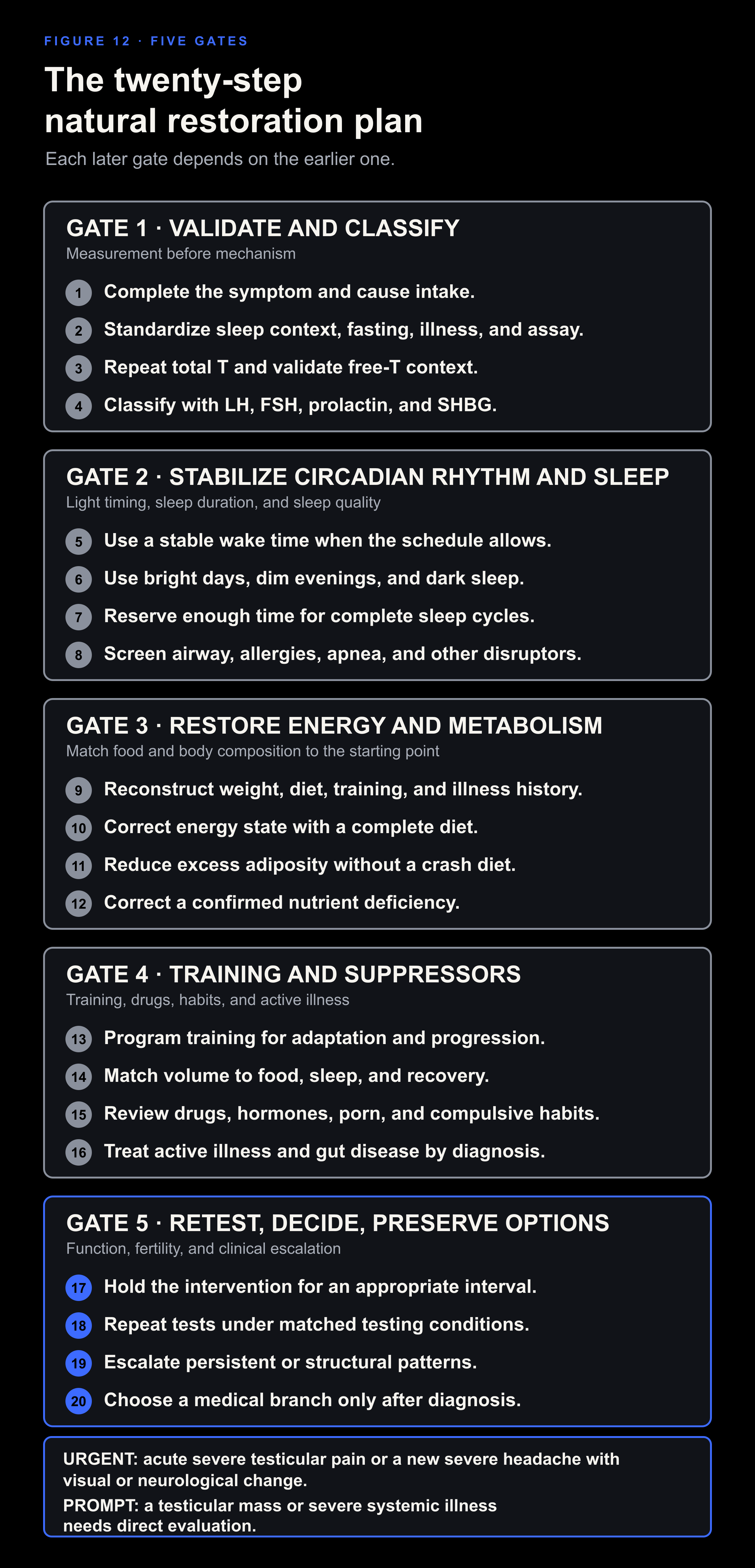
Figure 12. Each later intervention depends on a credible result at the earlier gate.
14. Daily and weekly practice
The plan works through repeatable anchors. Use the schedule that fits the diagnosed problem and the person’s real sleep phase.
The daily routine
This routine begins after urgent causes have been screened. Adjust the clock to the person’s actual sleep schedule. The anchors matter more than a universal bedtime.
After waking
Record wake time and morning state when sleep is an active problem.
Get outdoor light when feasible. Keep the timing consistent with the intended circadian phase.
Eat according to the diagnosed energy state. A man recovering from underfeeding needs enough food. A man reducing excess fat needs a sustainable deficit.
Take prescribed treatment as directed. Do not add a hormone or booster to the routine without a defined indication.
During the day
Eat enough complete protein and total energy for the plan. Set carbohydrate and fat by tolerance, activity, body composition, and clinical context.
Train only on scheduled days. Use progressive work that can be recovered from.
Keep caffeine early enough that sleep remains intact.[25]
Use a bright daytime environment. A dim day followed by a bright screen at night weakens the day-night contrast.
Before sleep
Dim light and reduce bright screens near the face.
Leave enough sleep opportunity for complete cycles rather than forcing one fixed sleep time.
Manage the known disruptor. This may be congestion, apnea treatment, pain, reflux, heat, alcohol, restless legs, or noise.
Use saline rinse or a nasal strip only when it fits the nasal problem. Mouth tape never replaces an open airway or apnea care.[62][61][59]
Record only the outcomes needed for the current hypothesis. A long nightly scorecard will not improve adherence.
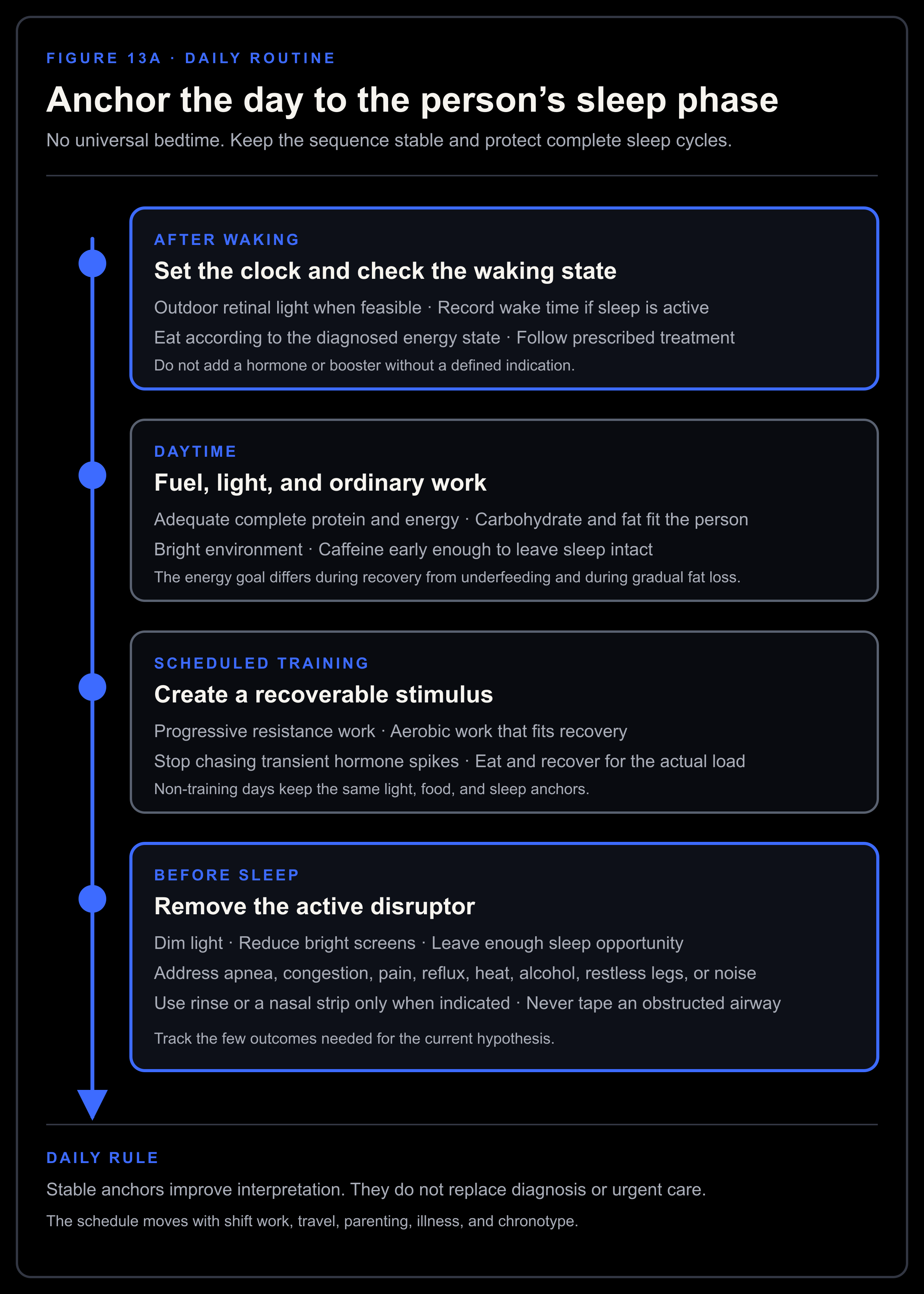
Figure 13A. Daily anchors create light contrast, adequate fuel, recoverable work, and protected sleep. The schedule follows the person’s sleep phase rather than a fixed clock time.
The weekly routine
Review trends once a week. Daily hormone interpretation creates noise.
Compare wake-time range, sleep opportunity, awakenings, and daytime sleepiness.
Review body-weight and waist direction without reacting to one reading.
Check whether food intake matched the energy goal and whether protein remained adequate.
Review training performance, total hard sets or endurance load, pain, and recovery.
Record libido, spontaneous or morning erections, and the main functional outcome chosen at baseline.
Check adherence to prescribed apnea, thyroid, allergy, or other cause-specific treatment.
Review alcohol, nicotine, drugs, hormones, supplements, porn, edging, and compulsive masturbation.
Adjust one active variable when possible. Keep the rest stable long enough to learn from the change.
Escalate new red flags, persistent high prolactin, fertility concerns, or a worsening laboratory pattern.
Laboratory testing belongs at the interval required by the biology. It does not belong on a weekly schedule. Semen recovery, fat loss, thyroid treatment, and recovery after AAS follow different timelines.
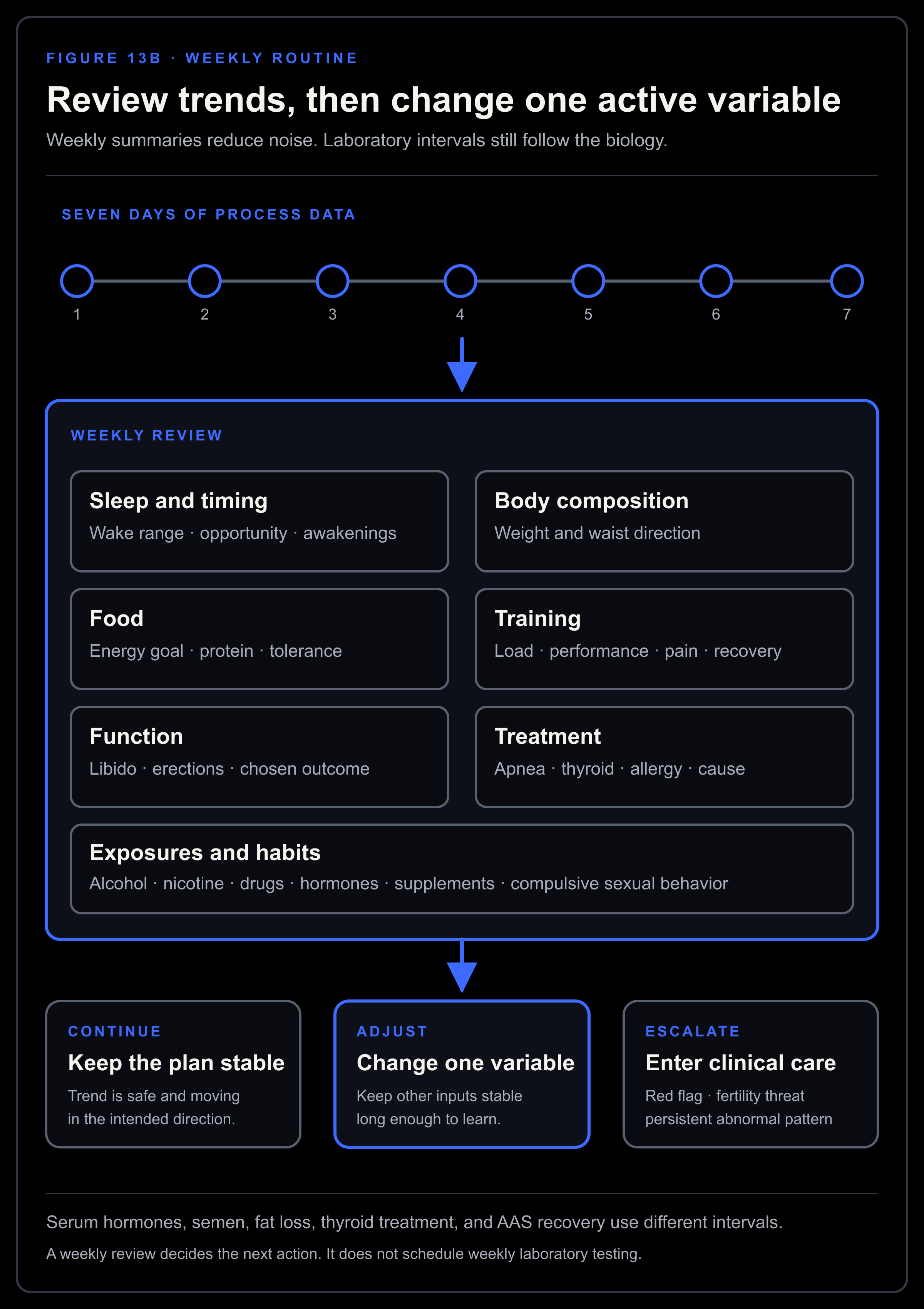
Figure 13B. The weekly review compares trends, checks the main limiting layer, and changes one variable before the next review.
15. Monitor the result
Choose outcomes before changing the plan
Keep the record small enough to use consistently:
Sleep timing, duration, awakenings, and morning state.
Daytime sleepiness.
Libido and frequency of spontaneous or morning erections.
Erectile reliability in the relevant context.
Training performance and recovery.
Body weight and waist trajectory.
Mood or validated symptom measures when appropriate.
Semen parameters when fertility is the target.
Total testosterone, SHBG or free testosterone, LH, FSH, and cause-specific laboratories.
Summarize the record weekly. Sexual function, mood, and sleep fluctuate too much for one day to carry much meaning.
Separate process measures from outcome measures
Process measures show whether the action happened. Wake-time consistency, CPAP use, food intake, training volume, and weight trend belong here. Outcome measures show whether the target changed, such as testosterone, sleepiness, libido, sperm concentration, strength, or waist circumference.
Perfect adherence can still test the wrong idea.
Match the reassessment interval to the biology
Light timing may help within days or weeks. Recovery from underfeeding takes longer, and fat loss unfolds over months. Recovery after steroid exposure can be slow and uncertain. Sperm production also changes more slowly than a serum hormone result.
Keep measurement conditions comparable
A higher result after better sleep may partly reflect better sampling conditions, which itself shows that context mattered.
With every clinically important test, record wake time, fasting state, illness, training, medication timing, and androgen exposure. Use the same reliable laboratory when possible so the comparison remains meaningful.
Define success at several levels
Success can include:
A valid laboratory pattern moving toward normal.
Improvement in the symptom that justified the work.
Preservation of fertility goals.
Removal or control of the upstream cause.
A plan that can be sustained without escalating risk.
Partial success still carries information. Better sleep with unchanged testosterone points toward another limiting layer. Higher testosterone with unchanged fatigue shifts attention toward another cause of the fatigue.
Define failure before it happens
Failure means poor adherence, an unusable measurement, no result after enough time, adverse effects, worsening fertility, or discovery of a structural cause. Define it before starting.
The reason for failure determines the next move:
Fix adherence when exposure was inadequate.
Improve measurement when the comparison was invalid.
Extend time when the biology is slow and the direction is safe.
Change the hypothesis when the intervention was adequate and the outcome did not move.
Escalate care when the pattern suggests pituitary, testicular, systemic, or medication-related disease.
Each intervention should test one layer of the model.
16. Pattern-based troubleshooting
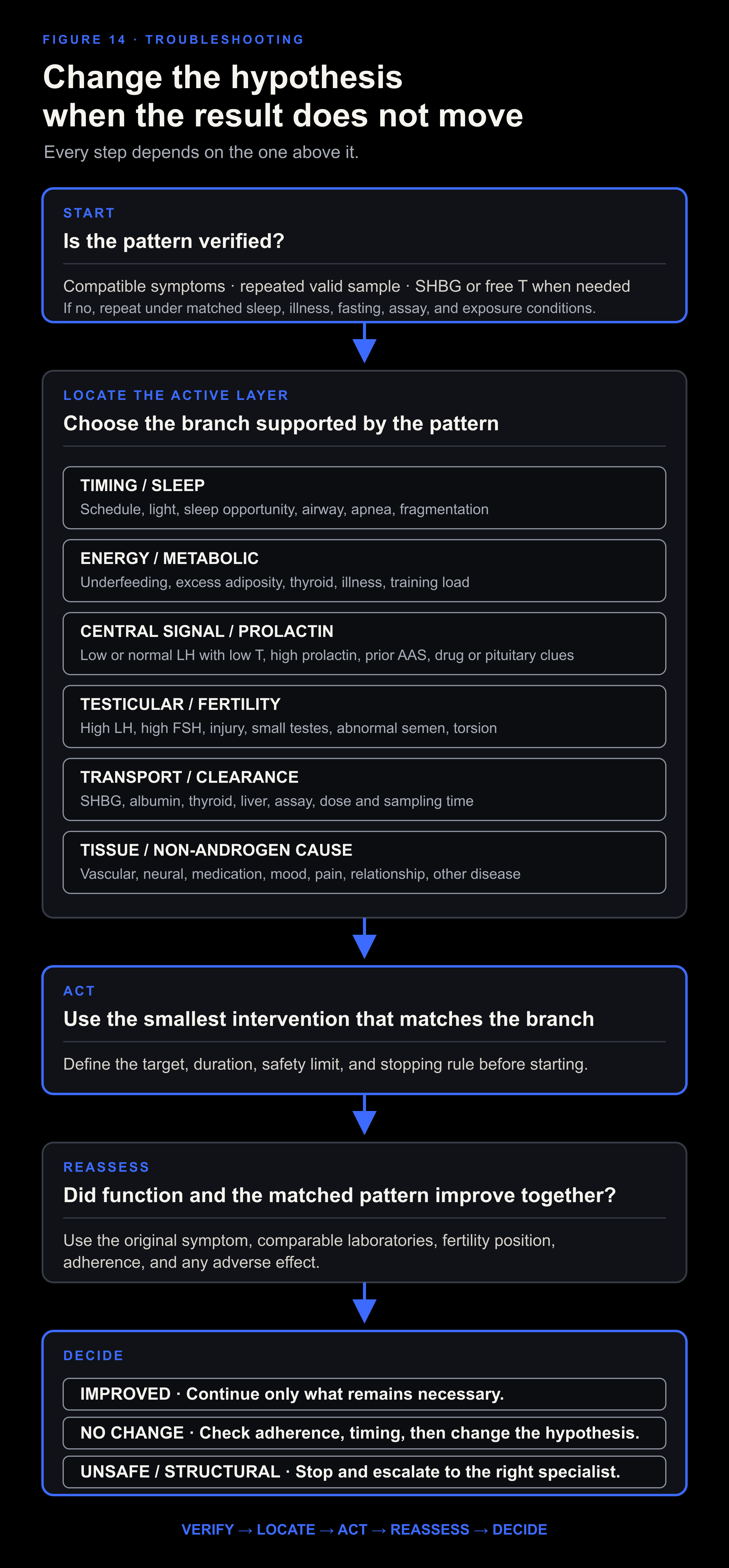
Figure 14. Troubleshooting begins with the pattern. Persistent symptoms with normal, valid androgen measurements require a broader diagnostic investigation.
Pattern A: low testosterone after poor sleep
First determine whether the result followed one unusual night or reflects a chronic pattern.
After an unusual night, restore the usual schedule and repeat the test. For a chronic pattern, distinguish among:
Insufficient opportunity.
Delayed or irregular circadian timing.
Insomnia.
OSA or another sleep disorder.
Caffeine, alcohol, nicotine, pain, reflux, or medication.
Shift work.
Treat the identified sleep problem, then repeat under representative conditions. High LH, markedly elevated prolactin, or structural clues point beyond sleep and need their own evaluation.
Pattern B: low total testosterone with obesity
First read the binding context. Measure SHBG, assess free testosterone, and add LH and FSH.
Low SHBG with preserved free testosterone shifts priority toward metabolic health and the symptom pattern. When both total and free testosterone are low alongside low or normal LH, functional central suppression becomes more likely, though pituitary and medication causes still need review. High LH means obesity alone cannot explain the primary pattern.
Screen for OSA. Review alcohol, opioids, diabetes, liver disease, and prior androgen use. Use a sustainable weight-loss plan and retest.
Pattern C: low testosterone during a cut
Reconstruct the deficit. Review its size and duration, the change in body fat, training load, sleep, illness, and dietary rigidity. Reduce the combined stress by raising intake, lowering expenditure, or doing both.
Preserve protein and restore enough carbohydrate and fat to make the diet complete. Give body weight, sleep, libido, and training performance time to recover before repeating the test.
Persistent high LH or a result that predates the cut points toward a different layer.
Pattern D: low testosterone with high LH
Treat this as a testicular pattern until the workup says otherwise. Confirm the result, include FSH, and arrange clinical evaluation. Review testicular history, fertility, infection, torsion, trauma, chemotherapy, radiation, genetic conditions, and examination findings.
Lifestyle can support general health while the testicular cause is being diagnosed.
Pattern E: low testosterone with low LH and high prolactin
Confirm prolactin and find the cause. Repeat it under appropriate conditions while reviewing medications, thyroid status, renal function, and symptoms. Marked or persistent elevation requires a search for the cause. Headache, visual change, galactorrhea, or other pituitary deficits make that search urgent.
Dopamine agonists require medical diagnosis and management.
Pattern F: normal testosterone with persistent symptoms
Confirm the androgen interpretation, then widen the diagnosis. First check free testosterone and SHBG.
For fatigue, consider sleep disorders, anemia, thyroid disease, depression, infection, cardiopulmonary disease, medication, underfueling, and overtraining.
For erectile dysfunction, consider vascular risk, blood pressure, diabetes, medication, pornography or stimulation patterns, anxiety, pelvic or neurological factors, and relationship context.
For low libido, consider mood, stress, medication, sleep, relationship factors, pain, prolactin, thyroid status, and estradiol context.
For poor training response, examine programming, technique, volume, food, protein, sleep, illness, and time.
A normal, valid androgen profile redirects the investigation without dismissing the symptoms.
Pattern G: high testosterone with suppressed LH and fertility concerns
Protect fertility before chasing the serum value. Document every androgen, SARM, prohormone, hCG, and fertility-drug exposure. Obtain a semen analysis and reproductive evaluation. Recovery after anabolic use varies and can be prolonged.
An internet post-cycle therapy protocol cannot account for the exposure, time course, testicular function, fertility timeline, and medical risks that should determine the plan.
Pattern H: numbers improve and function does not
Return to the original symptom. Reconsider whether the first attribution was correct.
An improved testosterone result settles only the androgen measurement. Persistent sleepiness, erectile dysfunction, depression, pain, infertility, or poor performance returns the investigation to the cause of that outcome.
Recognizing that mismatch prevents an endless sequence of higher doses and narrower explanations.
17. Where the natural approach ends
This article stops at endogenous production.
Sleep, food, training, body composition, thyroid care, airway treatment, and exposure reduction cannot rebuild destroyed testicular tissue or remove a pituitary tumor. Nor can they reverse every effect of chemotherapy, genetic disease, torsion, or prolonged steroid exposure.
The first article named thyroid hormone, progesterone, dopamine agonists, aromatase inhibitors, and other drugs. Their mechanisms belong in the map. The default natural protocol excludes them. Each drug needs a diagnosis, target, monitoring plan, fertility decision, and stopping rule.
Testosterone replacement therapy supplies the final hormone. It can normalize serum testosterone and improve selected outcomes in confirmed hypogonadism. It suppresses GnRH, LH, FSH, intratesticular testosterone, and sperm production. It is rational when clinically important androgen deficiency is confirmed and endogenous recovery is unavailable or inadequate. Apnea, pituitary disease, underfeeding, medication effects, and testicular disease remain separate treatment targets.[200][40]
hCG activates the LH receptor on Leydig cells. It can support intratesticular testosterone during gonadotropin suppression and is used in selected fertility and secondary-hypogonadism settings. Its action requires responsive Leydig cells and leaves FSH-dependent Sertoli function as a separate branch. Estradiol, symptoms, semen, and testicular response require monitoring.[200]
Clomiphene blocks estrogen feedback centrally and can raise endogenous LH, FSH, and testosterone when the hypothalamus, pituitary, and testes remain responsive. It is a mixture of enclomiphene and zuclomiphene and is used off label in men. It may preserve fertility better than testosterone, but semen responses vary and can worsen in a minority. It is a poor fit for primary testicular failure or a structural pituitary cause.[245]
Enclomiphene is the trans isomer of clomiphene. Trials in men with secondary hypogonadism found higher testosterone, LH, and FSH with preserved sperm counts compared with testosterone gel.[246] That is drug-driven stimulation of an intact axis. Continued sleep, obesity, medication, or pituitary causes can remain unresolved. Approval and availability vary by jurisdiction.
Enclomiphene should not be called a “side-effect-free natural testosterone booster.” It is a selective estrogen-receptor modulator that changes feedback. It belongs in a defined medical branch with monitoring.
Aromatase inhibitors reduce conversion of testosterone to estradiol and weaken estrogen feedback. In selected infertile or hypoandrogenic men with an appropriate testosterone-to-estradiol pattern, they can raise LH, FSH, testosterone, and some semen measures.[247] Estradiol is still required for bone, sexual function, and body composition. Broad suppression can create a new problem and does not treat every cause of high estradiol.
Dopamine agonists lower prolactin and can restore gonadal signaling when a defined hyperprolactinemic disorder is the cause. They are not treatments for pornography, a brief post-orgasm prolactin rise, or a vague low-dopamine theory. Pituitary imaging, medication review, adverse effects, and long-term management belong to the clinical branch.[67]
Thyroid hormone treats diagnosed thyroid disease. Restoring euthyroidism can improve SHBG, prolactin, sexual function, and testicular output when hypothyroidism is the limiting layer.[189][190] T3 is one medical option within thyroid care. Excess exposure can disturb rhythm, sleep, muscle, bone, and the heart. Thyroid hormone has no routine role as a testosterone booster in a euthyroid man.
Pregnenolone is the first steroid after cholesterol. It can feed progesterone, DHEA, corticosteroid, and neurosteroid branches. Taking it does not determine which branch receives the substrate, and it does not guarantee more testosterone.[85]
Progesterone is a normal male steroid intermediate and neurosteroid precursor. Sufficient exposure can suppress LH, FSH, and testosterone. Its use therefore requires a defined purpose and monitoring of the axis and fertility.[243]
DHT supplies a potent nonaromatizable androgen signal. It can act at target tissues while suppressing LH, testosterone, and estradiol. A long trial preserved many sexual measures while reducing spinal bone density.[244][224] DHT cannot supply estrogen-dependent effects or preserve endogenous fertility.
DHEA is an exogenous steroid precursor. Tissue enzymes can convert it toward androgens or estrogens. Controlled trials in men show no reliable restoration of testosterone, sexual function, strength, or broader clinical outcomes.[248][249] It still changes downstream exposure and laboratory interpretation.
Post-cycle drug stacks combine hCG, selective estrogen-receptor modulators, aromatase inhibitors, and sometimes other hormones after AAS or testosterone exposure. Each component pushes a different branch. The HAARLEM cohort observed self-selected post-cycle therapy rather than testing it as a randomized intervention.[240] Recovery depends on the compounds, exposure, baseline testicular capacity, time, and fertility goal. A stack can also obscure whether autonomous function has returned.
The separate HRT, ICT and PCT framework defines HRT as long-term hormone support, ICT as support during active AAS exposure, and PCT as assessment and recovery support after exposure has cleared. This guide does not tell readers to progress through them. A fully natural plan stops before hormone or fertility drugs. When a medical branch becomes necessary, define the failed layer and protect fertility before treatment begins.
The final decision rule
The first Testosterone Kabbalah had the right instinct. Testosterone belongs inside a network. The harder lesson is that every blood value leaves something out. Serum testosterone cannot show intratesticular testosterone, local DHT or estradiol, receptor action, fertility, or the cause of a symptom.
Start with a result worth interpreting. Read LH, FSH, SHBG, free testosterone when needed, prolactin, and the clinical context beside it. The pattern will usually point toward sleep, energy, metabolism, thyroid, medication, pituitary signaling, testicular function, transport, conversion, or a different cause of the symptom.
Natural restoration has a precise meaning. Repair the limiting branch while it can still recover. Retest under matched conditions. Stop self-experimenting when the pattern calls for medical treatment.
Protect fertility before changing the axis. The number should serve the diagnosis.
References
Ananda S. Prasad et al. “Zinc Status and Serum Testosterone Levels of Healthy Adults.” Nutrition 12, no. 5 (1996): 344-348. DOI: 10.1016/S0899-9007(96)80058-X.
Stephen J. Smith, Adrian L. Lopresti, Shaun Y. M. Teo, and Timothy J. Fairchild. “Examining the Effects of Herbs on Testosterone Concentrations in Men: A Systematic Review.” Advances in Nutrition 12, no. 3 (2021): 744-765. DOI: 10.1093/advances/nmaa134.
Alexandre Morgado et al. “Do ‘Testosterone Boosters’ Really Increase Serum Total Testosterone? A Systematic Review.” International Journal of Impotence Research 36, no. 4 (2024): 348-364. DOI: 10.1038/s41443-023-00763-9.
Musa T. Yakubu, Musbau A. Akanji, and Adenike T. Oladiji. “Aphrodisiac Potentials of the Aqueous Extract of Fadogia agrestis Stem in Male Albino Rats.” Asian Journal of Andrology 7, no. 4 (2005): 399-404. DOI: 10.1111/j.1745-7262.2005.00052.x.
Musa T. Yakubu, Musbau A. Akanji, and Adenike T. Oladiji. “Effects of Oral Administration of Aqueous Extract of Fadogia agrestis Stem on Some Testicular Function Indices of Male Rats.” Journal of Ethnopharmacology 115, no. 2 (2008): 288-292. DOI: 10.1016/j.jep.2007.10.004.
Musa T. Yakubu and Anthony J. Afolayan. “Reproductive Toxicologic Evaluations of Bulbine natalensis Baker Stem Extract in Albino Rats.” Theriogenology 72, no. 3 (2009): 322-332. DOI: 10.1016/j.theriogenology.2009.01.026.
Anthony J. Afolayan and Musa T. Yakubu. “Effect of Bulbine natalensis Baker Stem Extract on the Functional Indices and Histology of the Liver and Kidney of Male Wistar Rats.” Journal of Medicinal Food 12, no. 4 (2009): 814-820. DOI: 10.1089/jmf.2008.0221.
Vikas Sharma, Mayank Thakur, Nagendra S. Chauhan, and Vinod Kumar Dixit. “Effects of Petroleum Ether Extract of Anacyclus pyrethrum DC. on Sexual Behavior in Male Rats.” Journal of Chinese Integrative Medicine 8, no. 8 (2010): 767-773. DOI: 10.3736/jcim20100807.
V. K. Neychev and V. I. Mitev. “The Aphrodisiac Herb Tribulus terrestris Does Not Influence the Androgen Production in Young Men.” Journal of Ethnopharmacology 101, nos. 1-3 (2005): 319-323. DOI: 10.1016/j.jep.2005.05.017.
Carlos A. Santos Jr. et al. “Tribulus terrestris versus Placebo in the Treatment of Erectile Dysfunction: A Prospective, Randomized, Double-Blind Study.” Actas Urológicas Españolas 38, no. 4 (2014): 244-248. DOI: 10.1016/j.acuro.2013.09.014.
José de Oliveira Vilar Neto et al. “Effects of Tribulus terrestris Supplementation on Erectile Dysfunction and Testosterone Levels in Men: A Systematic Review of Clinical Trials.” Nutrients 17, no. 7 (2025): 1275. DOI: 10.3390/nu17071275.
W. Cherdshewasart and N. Nimsakul. “Clinical Trial of Butea superba, an Alternative Herbal Treatment for Erectile Dysfunction.” Asian Journal of Andrology 5, no. 3 (2003): 243-246.
Jeff R. Cortés-González, Jorge A. Arratia-Maqueo, Lauro S. Gómez-Guerra, and Anders R. Holmberg. “The Use of Butea superba (Roxb.) Compared to Sildenafil for Treating Erectile Dysfunction.” BJU International 105, no. 2 (2010): 225-228. DOI: 10.1111/j.1464-410X.2009.08748.x.
Kamon Chaiyasit and Viroj Wiwanitkit. “Hyperandrogenemia Due to Ingestion of Butea superba.” Indian Journal of Endocrinology and Metabolism 16, no. 3 (2012): 485-486. DOI: 10.4103/2230-8210.95756.
Kristian Leisegang, Renata Finelli, Suresh C. Sikka, and Manesh Kumar Panner Selvam. “Eurycoma longifolia (Jack) Improves Serum Total Testosterone in Men: A Systematic Review and Meta-Analysis of Clinical Trials.” Medicina 58, no. 8 (2022): 1047. DOI: 10.3390/medicina58081047.
Sasikala M. Chinnappan et al. “Effect of Eurycoma longifolia Standardised Aqueous Root Extract, Physta, on Testosterone Levels and Quality of Life in Ageing Male Subjects: A Randomised, Double-Blind, Placebo-Controlled Multicentre Study.” Food & Nutrition Research 65 (2021): 5647. DOI: 10.29219/fnr.v65.5647.
Kai Quin Chan, Claire Stewart, Neil Chester, Sareena H. Hamzah, and Ashril Yusof. “The Effect of Eurycoma longifolia on the Regulation of Reproductive Hormones in Young Males.” Andrologia 53, no. 4 (2021): e14001. DOI: 10.1111/and.14001.
Adrian L. Lopresti, Peter D. Drummond, and Stephen J. Smith. “A Randomized, Double-Blind, Placebo-Controlled, Crossover Study Examining the Hormonal and Vitality Effects of Ashwagandha in Aging, Overweight Males.” American Journal of Men’s Health 13, no. 2 (2019): 1557988319835985. DOI: 10.1177/1557988319835985.
Sindre Lee-Ødegård, Thomas E. Gundersen, and Christian A. Drevon. “Effect of a Plant Extract of Fenugreek on Testosterone in Blood Plasma and Saliva in a Double-Blind Randomized Controlled Intervention Study.” PLOS ONE 19, no. 9 (2024): e0310170. DOI: 10.1371/journal.pone.0310170.
S. Pandit et al. “Clinical Evaluation of Purified Shilajit on Testosterone Levels in Healthy Volunteers.” Andrologia 48, no. 5 (2016): 570-575. DOI: 10.1111/and.12482.
Elisabeth Lerchbaum et al. “Vitamin D and Testosterone in Healthy Men: A Randomized Controlled Trial.” Journal of Clinical Endocrinology & Metabolism 102, no. 11 (2017): 4292-4302. DOI: 10.1210/jc.2017-01428.
Alexander M. Sidlak et al. “Acute Salicylate Toxicity: A Narrative Review for Emergency Clinicians.” Cureus 17, no. 9 (2025): e91505. DOI: 10.7759/cureus.91505.
Su Ling Young and Gus Gazzard. “The Adverse Effects of Oral Niacin/Nicotinamide: An Overview of Reviews.” Eye 39, no. 16 (2025): 2852-2859. DOI: 10.1038/s41433-025-04027-2.
M. J. Stampfer et al. “Vitamin E Supplementation Effect on Human Platelet Function, Arachidonic Acid Metabolism, and Plasma Prostacyclin Levels.” American Journal of Clinical Nutrition 47, no. 4 (1988): 700-706. DOI: 10.1093/ajcn/47.4.700.
Carissa Gardiner et al. “The Effect of Caffeine on Subsequent Sleep: A Systematic Review and Meta-Analysis.” Sleep Medicine Reviews 69 (2023): 101764. DOI: 10.1016/j.smrv.2023.101764.
Geoffrey W. Melville, Jason C. Siegler, and Paul W. M. Marshall. “The Effects of D-Aspartic Acid Supplementation in Resistance-Trained Men over a Three-Month Training Period: A Randomised Controlled Trial.” PLOS ONE 12, no. 8 (2017): e0182630. DOI: 10.1371/journal.pone.0182630.
Joel S. Finkelstein et al. “Gonadal Steroids and Body Composition, Strength, and Sexual Function in Men.” New England Journal of Medicine 369, no. 11 (2013): 1011-1022. DOI: 10.1056/NEJMoa1206168.
Dongho Shin et al. “Efficacy and Safety of Maca in Patients with Symptoms of Late-Onset Hypogonadism: A Randomized, Double-Blind, Placebo-Controlled Clinical Trial.” World Journal of Men’s Health 41, no. 3 (2023): 692-700. DOI: 10.5534/wjmh.220112.
Johann van der Merwe, Naomi E. Brooks, and Kathryn H. Myburgh. “Three Weeks of Creatine Monohydrate Supplementation Affects Dihydrotestosterone to Testosterone Ratio in College-Aged Rugby Players.” Clinical Journal of Sport Medicine 19, no. 5 (2009): 399-404. DOI: 10.1097/JSM.0b013e3181b8b52f.
Gregory D. M. Potter et al. “Effects of Exercise Training on Resting Testosterone Concentrations in Insufficiently Active Men: A Systematic Review and Meta-Analysis.” Journal of Strength and Conditioning Research 36, no. 12 (2022): 3521-3528. DOI: 10.1519/JSC.0000000000004146.
Daniel W. D. West et al. “Resistance Exercise-Induced Increases in Putative Anabolic Hormones Do Not Enhance Muscle Protein Synthesis or Intracellular Signalling in Young Men.” Journal of Physiology 587, no. 21 (2009): 5239-5247. DOI: 10.1113/jphysiol.2009.177220.
Tillmann H. Krüger et al. “Orgasm-Induced Prolactin Secretion: Feedback Control of Sexual Drive?” Neuroscience & Biobehavioral Reviews 26, no. 1 (2002): 31-44. DOI: 10.1016/S0149-7634(01)00036-7.
Tillmann H. Krüger et al. “Specificity of the Neuroendocrine Response to Orgasm during Sexual Arousal in Men.” Journal of Endocrinology 177, no. 1 (2003): 57-64. DOI: 10.1677/joe.0.1770057.
Alonso Fernández-Guasti and Gabriela Rodríguez-Manzo. “Pharmacological and Physiological Aspects of Sexual Exhaustion in Male Rats.” Scandinavian Journal of Psychology 44, no. 3 (2003): 257-263. DOI: 10.1111/1467-9450.00343.
Bryan V. Phillips-Farfán and Alonso Fernández-Guasti. “Endocrine, Neural and Pharmacological Aspects of Sexual Satiety in Male Rats.” Neuroscience & Biobehavioral Reviews 33, no. 3 (2009): 442-455. DOI: 10.1016/j.neubiorev.2008.11.003.
Zisis Zacharopoulos et al. “Pornography Consumption and Male Sexual Dysfunction: A Systematic Review.” Advances in Experimental Medicine and Biology 1487 (2026): 297-304. DOI: 10.1007/978-3-032-03398-7_29.
Beáta Bőthe et al. “Are Sexual Functioning Problems Associated with Frequent Pornography Use and/or Problematic Pornography Use? Results from a Large Community Survey Including Males and Females.” Addictive Behaviors 112 (2021): 106603. DOI: 10.1016/j.addbeh.2020.106603.
Neil Campbell et al. “Adulteration of Purported Herbal and Natural Sexual Performance Enhancement Dietary Supplements with Synthetic Phosphodiesterase Type 5 Inhibitors.” Journal of Sexual Medicine 10, no. 7 (2013): 1842-1849. DOI: 10.1111/jsm.12172.
Ryan M. Van Wagoner et al. “Chemical Composition and Labeling of Substances Marketed as Selective Androgen Receptor Modulators and Sold via the Internet.” JAMA 318, no. 20 (2017): 2004-2010. DOI: 10.1001/jama.2017.17069.
Shalender Bhasin et al. “Testosterone Therapy in Men with Hypogonadism: An Endocrine Society Clinical Practice Guideline.” Journal of Clinical Endocrinology & Metabolism 103, no. 5 (2018): 1715-1744. DOI: 10.1210/jc.2018-00229.
John E. Morley et al. “Validation of a Screening Questionnaire for Androgen Deficiency in Aging Males.” Metabolism 49, no. 9 (2000): 1239-1242. DOI: 10.1053/meta.2000.8625.
Sat Bir S. Khalsa et al. “A Phase Response Curve to Single Bright Light Pulses in Human Subjects.” Journal of Physiology 549, no. 3 (2003): 945-952. DOI: 10.1113/jphysiol.2003.040477.
Joshua J. Gooley et al. “Exposure to Room Light before Bedtime Suppresses Melatonin Onset and Shortens Melatonin Duration in Humans.” Journal of Clinical Endocrinology & Metabolism 96, no. 3 (2011): E463-E472. DOI: 10.1210/jc.2010-2098.
Gary A. Wittert. “The Relationship Between Sleep Disorders and Testosterone in Men.” Asian Journal of Andrology 16, no. 2 (2014): 262-265. DOI: 10.4103/1008-682X.122586.
Rafael Luboshitzky et al. “Relationship Between Rapid Eye Movement Sleep and Testosterone Secretion in Normal Men.” Journal of Andrology 20, no. 6 (1999): 731-737.
Rafael Luboshitzky et al. “Disruption of Nocturnal Testosterone Rhythm by Sleep Fragmentation in Normal Men.” Journal of Clinical Endocrinology & Metabolism 86, no. 3 (2001): 1134-1139. DOI: 10.1210/jcem.86.3.7296.
Yulia V. Ukraintseva et al. “Slow-Wave Sleep and Androgens: Selective Slow-Wave Sleep Suppression Affects Testosterone and 17α-Hydroxyprogesterone Secretion.” Sleep Medicine 48 (2018): 117-126. DOI: 10.1016/j.sleep.2018.04.012.
Long Su et al. “Effect of Partial and Total Sleep Deprivation on Serum Testosterone in Healthy Males: A Systematic Review and Meta-Analysis.” Sleep Medicine 88 (2021): 267-273. DOI: 10.1016/j.sleep.2021.10.031.
Rachel Leproult and Eve Van Cauter. “Effect of 1 Week of Sleep Restriction on Testosterone Levels in Young Healthy Men.” JAMA 305, no. 21 (2011): 2173-2174. DOI: 10.1001/jama.2011.710.
Brittany N. Harding et al. “Changes in Melatonin and Sex Steroid Hormone Production among Men as a Result of Rotating Night Shift Work: The HORMONIT Study.” Scandinavian Journal of Work, Environment & Health 48, no. 1 (2022): 41-51. DOI: 10.5271/sjweh.3991.
David I. Spratt et al. “Neuroendocrine-Gonadal Axis in Men: Frequent Sampling of LH, FSH, and Testosterone.” American Journal of Physiology 254, no. 5 Pt 1 (1988): E658-E666. DOI: 10.1152/ajpendo.1988.254.5.E658.
Claude Gronfier and Gabrielle Brandenberger. “Ultradian Rhythms in Pituitary and Adrenal Hormones: Their Relations to Sleep.” Sleep Medicine Reviews 2, no. 1 (1998): 17-29. DOI: 10.1016/S1087-0792(98)90051-X.
Li-Qiang Qin et al. “The Effects of Nocturnal Life on the Circadian Patterns of Sex Hormones in Young Healthy Men.” Journal of Health Science 51, no. 1 (2005): 33-40. DOI: 10.1248/jhs.51.33.
Helen J. Burgess, Victoria L. Revell, and Charmane I. Eastman. “A Three Pulse Phase Response Curve to Three Milligrams of Melatonin in Humans.” Journal of Physiology 586, no. 2 (2008): 639-647. DOI: 10.1113/jphysiol.2007.143180.
Alfred J. Lewy et al. “The Human Phase Response Curve to Melatonin Is about 12 Hours Out of Phase with the Phase Response Curve to Light.” Chronobiology International 15, no. 1 (1998): 71-83. DOI: 10.3109/07420529808998671.
Bhanu Prakash Kolla et al. “The Impact of Alcohol on Breathing Parameters during Sleep: A Systematic Review and Meta-Analysis.” Sleep Medicine Reviews 42 (2018): 59-67. DOI: 10.1016/j.smrv.2018.05.007.
John W. Winkelman et al. “Treatment of Restless Legs Syndrome and Periodic Limb Movement Disorder: An American Academy of Sleep Medicine Clinical Practice Guideline.” Journal of Clinical Sleep Medicine 21, no. 1 (2025): 137-152. DOI: 10.5664/jcsm.11390.
Long Su et al. “Association between Obstructive Sleep Apnea and Male Serum Testosterone: A Systematic Review and Meta-Analysis.” Andrology 10, no. 2 (2022): 223-231. DOI: 10.1111/andr.13111.
Kristina E. Hermelingmeier et al. “Nasal Irrigation as an Adjunctive Treatment in Allergic Rhinitis: A Systematic Review and Meta-Analysis.” American Journal of Rhinology & Allergy 26, no. 5 (2012): e119-e125. DOI: 10.2500/ajra.2012.26.3787.
Jonathan S. Yoder et al. “Primary Amebic Meningoencephalitis Deaths Associated with Sinus Irrigation Using Contaminated Tap Water.” Clinical Infectious Diseases 55, no. 9 (2012): e79-e85. DOI: 10.1093/cid/cis626.
Macario Camacho et al. “Nasal Dilators for Snoring and OSA: A Systematic Review and Meta-Analysis.” Pulmonary Medicine 2016 (2016): 4841310. DOI: 10.1155/2016/4841310.
Jared Rhee et al. “Breaking Social Media Fads and Uncovering the Safety and Efficacy of Mouth Taping in Patients with Mouth Breathing, Sleep Disordered Breathing, or Obstructive Sleep Apnea: A Systematic Review.” PLOS ONE 20, no. 5 (2025): e0323643. DOI: 10.1371/journal.pone.0323643.
Alfredo Cignarelli et al. “Effects of CPAP on Testosterone Levels in Patients with Obstructive Sleep Apnea: A Meta-Analysis Study.” Frontiers in Endocrinology 10 (2019): 551. DOI: 10.3389/fendo.2019.00551.
Timothy M. Brown. “Melanopic Illuminance Defines the Magnitude of Human Circadian Light Responses under a Wide Range of Conditions.” Journal of Pineal Research 69, no. 1 (2020): e12655. DOI: 10.1111/jpi.12655.
P. Michael Iuvone et al. “Light Stimulates Tyrosine Hydroxylase Activity and Dopamine Synthesis in Retinal Amacrine Neurons.” Science 202, no. 4370 (1978): 901-902. DOI: 10.1126/science.30997.
Georgi Dinkov. “Lack of Light Exposure May Be the Main Cause of the Myopia Boom.” Official website, October 10, 2019.
Shlomo Melmed et al. “Diagnosis and Treatment of Hyperprolactinemia: An Endocrine Society Clinical Practice Guideline.” Journal of Clinical Endocrinology & Metabolism 96, no. 2 (2011): 273-288. DOI: 10.1210/jc.2010-1692.
M. Mikhaylova et al. “The Effects of Ladasten on Dopaminergic Neurotransmission and Hippocampal Synaptic Plasticity in Rats.” Neuropharmacology 53, no. 5 (2007): 601-608. DOI: 10.1016/j.neuropharm.2007.07.001.
T. V. Grekhova et al. “Effect of Bromantane, a New Immunostimulating Agent with Psychostimulating Activity, on the Release and Metabolism of Dopamine in the Striatum of Freely Moving Rats: A Microdialysis Study.” Bulletin of Experimental Biology and Medicine 119, no. 3 (1995): 294-296. DOI: 10.1007/BF02445840.
Yu. V. Vakhitova et al. “cDNA Macroarray Analysis of Gene Expression Changes in Rat Brain after a Single Administration of a 2-Aminoadamantane Derivative.” Molecular Biology 39, no. 2 (2005): 244-252. DOI: 10.1007/s11008-005-0035-7.
Sergiy Oliynyk and Seikwan Oh. “The Pharmacology of Actoprotectors: Practical Application for Improvement of Mental and Physical Performance.” Biomolecules & Therapeutics 20, no. 5 (2012): 446-456. DOI: 10.4062/biomolther.2012.20.5.446.
Daniele Santi et al. “Seasonal Changes of Serum Gonadotropins and Testosterone in Men Revealed by a Large Data Set of Real-World Observations Over Nine Years.” Frontiers in Endocrinology 10 (2019): 914. DOI: 10.3389/fendo.2019.00914.
T. Zornitzki et al. “Seasonal Variation of Testosterone Levels in a Large Cohort of Men.” International Journal of Endocrinology 2022 (2022): 6093092. DOI: 10.1155/2022/6093092.
Laura M. Rossow et al. “Natural Bodybuilding Competition Preparation and Recovery: A 12-Month Case Study.” International Journal of Sports Physiology and Performance 8, no. 5 (2013): 582-592. DOI: 10.1123/ijspp.8.5.582.
Karsten Koehler et al. “Low Energy Availability in Exercising Men Is Associated with Reduced Leptin and Insulin but Not with Changes in Other Metabolic Hormones.” Journal of Sports Sciences 34, no. 20 (2016): 1921-1929. DOI: 10.1080/02640414.2016.1142109.
Ivan Jurov et al. “Reducing Energy Availability in Male Endurance Athletes: A Randomized Trial with a Three-Step Energy Reduction.” Journal of the International Society of Sports Nutrition 19, no. 1 (2022): 179-195. DOI: 10.1080/15502783.2022.2065111.
Alexiaa Sim, Hui Qing Tan, Yusuf Ali, and Stephen F. Burns. “Original Investigation: Manipulating Energy Availability in Male Endurance Runners: A Randomised Controlled Trial.” Applied Physiology, Nutrition, and Metabolism 49, no. 9 (2024): 1163-1174. DOI: 10.1139/apnm-2024-0037.
Joseph Whittaker and Kexin Wu. “Low-Fat Diets and Testosterone in Men: Systematic Review and Meta-Analysis of Intervention Studies.” Journal of Steroid Biochemistry and Molecular Biology 210 (2021): 105878. DOI: 10.1016/j.jsbmb.2021.105878.
Sahar Soltani et al. “The Effect of Low-Fat Diets Versus High-Fat Diet on Sex Hormones: A Systematic Review and Meta-Analysis of Randomized Controlled Trials.” Journal of Food Science 90, no. 5 (2025): e70266. DOI: 10.1111/1750-3841.70266.
Ray Peat. “Fatigue, Aging, and Recuperation.” RayPeat.com.
Ray Peat. “Fats and Degeneration.” 2009. RayPeat.com.
Ray Peat. “Gelatin, Stress, Longevity.” RayPeat.com.
Mathis Grossmann. “Hypogonadism and Male Obesity: Focus on Unresolved Questions.” Clinical Endocrinology 89, no. 1 (2018): 11-21. DOI: 10.1111/cen.13723.
Giovanni Corona et al. “Body Weight Loss Reverts Obesity-Associated Hypogonadotropic Hypogonadism: A Systematic Review and Meta-Analysis.” European Journal of Endocrinology 168, no. 6 (2013): 829-843. DOI: 10.1530/EJE-12-0955.
Walter L. Miller and Richard J. Auchus. “The Molecular Biology, Biochemistry, and Physiology of Human Steroidogenesis and Its Disorders.” Endocrine Reviews 32, no. 1 (2011): 81-151. DOI: 10.1210/er.2010-0013.
Barry R. Zirkin and Vassilios Papadopoulos. “Leydig Cells: Formation, Function, and Regulation.” Biology of Reproduction 99, no. 1 (2018): 101-111. DOI: 10.1093/biolre/ioy059.
Federico Pietrocola et al. “Acetyl Coenzyme A: A Central Metabolite and Second Messenger.” Cell Metabolism 21, no. 6 (2015): 805-821. DOI: 10.1016/j.cmet.2015.05.014.
Joseph L. Goldstein and Michael S. Brown. “Regulation of the Mevalonate Pathway.” Nature 343 (1990): 425-430. DOI: 10.1038/343425a0.
Niels A. Graudal, Thorbjørn Hubeck-Graudal, and Gesche Jürgens. “Effects of Low Sodium Diet versus High Sodium Diet on Blood Pressure, Renin, Aldosterone, Catecholamines, Cholesterol, and Triglyceride.” Cochrane Database of Systematic Reviews 12 (2020): CD004022. DOI: 10.1002/14651858.CD004022.pub5.
Ray Peat. “Salt, Energy, Metabolic Rate, and Longevity.” RayPeat.com.
Angela X. Chen et al. “Dietary Sodium Intake and Cortisol Measurements.” Clinical Endocrinology 93, no. 5 (2020): 539-545. DOI: 10.1111/cen.14262.
Derrick Lonsdale. “A Review of the Biochemistry, Metabolism and Clinical Benefits of Thiamin(e) and Its Derivatives.” Evidence-Based Complementary and Alternative Medicine 3, no. 1 (2006): 49-59. DOI: 10.1093/ecam/nek009.
Cristian Díaz-Muñoz et al. “Genetic Dissection of Stool Frequency Implicates Vitamin B1 Metabolism and Other Actionable Pathways in the Modulation of Gut Motility.” Gut (2026). DOI: 10.1136/gutjnl-2025-337059.
Georgi Dinkov. “Vitamin B1 (Thiamine) May Improve Gut Motility and Relieve Constipation/IBS.” Official website, May 24, 2026.
Georgi Dinkov. “Increased Fat Oxidation (FAO) Sufficient to Cause Senescence/Aging/Disease.” Official website, November 1, 2024.
Georgi Dinkov. “Low Energy Production (Glucose Metabolism) May Cause Andropause.” Official website, November 3, 2020.
Wataru Yamadera et al. “Glycine Ingestion Improves Subjective Sleep Quality in Human Volunteers, Correlating with Polysomnographic Changes.” Sleep and Biological Rhythms 5, no. 2 (2007): 126-131. DOI: 10.1111/j.1479-8425.2007.00262.x.
Amanda Bulman et al. “The Effects of L-Theanine Consumption on Sleep Outcomes: A Systematic Review and Meta-Analysis.” Sleep Medicine Reviews 81 (2025): 102076. DOI: 10.1016/j.smrv.2025.102076.
Eunsoo Moon et al. “Melatonergic Agents Influence the Sleep-Wake and Circadian Rhythms in Healthy and Psychiatric Participants: A Systematic Review and Meta-Analysis of Randomized Controlled Trials.” Neuropsychopharmacology 47, no. 8 (2022): 1523-1536. DOI: 10.1038/s41386-022-01278-5.
N. D. Goncharova, B. Kh. Khavinson, and B. A. Lapin. “Regulatory Effect of Epithalon on Production of Melatonin and Cortisol in Old Monkeys.” Bulletin of Experimental Biology and Medicine 131, no. 4 (2001): 394-396. DOI: 10.1023/A:1017928925177.
Szymon Kamil Araj et al. “Overview of Epitalon: Highly Bioactive Pineal Tetrapeptide with Promising Properties.” International Journal of Molecular Sciences 26, no. 6 (2025): 2691. DOI: 10.3390/ijms26062691.
S. Maria O’Kane et al. “Micronutrients, Iodine Status and Concentrations of Thyroid Hormones: A Systematic Review.” Nutrition Reviews 76, no. 6 (2018): 418-431. DOI: 10.1093/nutrit/nuy008.
Jessica Farebrother, Michael B. Zimmermann, and Maria Andersson. “Excess Iodine Intake: Sources, Assessment, and Effects on Thyroid Function.” Annals of the New York Academy of Sciences 1446, no. 1 (2019): 44-65. DOI: 10.1111/nyas.14041.
Camilla Bøgelund Larsen et al. “Selenium Supplementation and Placebo Are Equally Effective in Improving Quality of Life in Patients with Hypothyroidism.” European Thyroid Journal 13, no. 1 (2024). DOI: 10.1530/ETJ-23-0175.
Maurizio Nordio and Raffaella Pajalich. “Combined Treatment with Myo-Inositol and Selenium Ensures Euthyroidism in Subclinical Hypothyroidism Patients with Autoimmune Thyroiditis.” Journal of Thyroid Research 2013 (2013): 424163. DOI: 10.1155/2013/424163.
Doaa El Amrousy et al. “Lactoferrin for Iron-Deficiency Anemia in Children with Inflammatory Bowel Disease: A Clinical Trial.” Pediatric Research 92, no. 3 (2022): 762-766. DOI: 10.1038/s41390-022-02136-2.
Huma Bokkhim et al. “Physico-Chemical Properties of Different Forms of Bovine Lactoferrin.” Food Chemistry 141, no. 3 (2013): 3007-3013. DOI: 10.1016/j.foodchem.2013.05.139.
Dominik Kappeler et al. “Higher Bioavailability of Magnesium Citrate as Compared to Magnesium Oxide Shown by Evaluation of Urinary Excretion and Serum Levels after Single-Dose Administration in a Randomized Cross-Over Study.” BMC Nutrition 3 (2017): 7. DOI: 10.1186/s40795-016-0121-3.
Uwe Gröber et al. “Myth or Reality: Transdermal Magnesium?” Nutrients 9, no. 8 (2017): 813. DOI: 10.3390/nu9080813.
Arman Arab et al. “The Role of Magnesium in Sleep Health: A Systematic Review of Available Literature.” Biological Trace Element Research 201, no. 1 (2023): 121-128. DOI: 10.1007/s12011-022-03162-1.
Emma A. Vermeulen and Marc G. Vervloet. “Magnesium Administration in Chronic Kidney Disease.” Nutrients 15, no. 3 (2023): 547. DOI: 10.3390/nu15030547.
Asagi Ito et al. “Menaquinone-4 Enhances Testosterone Production in Rats and Testis-Derived Tumor Cells.” Lipids in Health and Disease 10 (2011): 158. DOI: 10.1186/1476-511X-10-158.
Stephen Schaffer, K. Takahashi, and Junichi Azuma. “Role of Osmoregulation in the Actions of Taurine.” Amino Acids 19, nos. 3-4 (2000): 527-546. DOI: 10.1007/s007260070004.
Vladimir Khavinson et al. “EDR Peptide: Possible Mechanism of Gene Expression and Protein Synthesis Regulation Involved in the Pathogenesis of Alzheimer’s Disease.” Molecules 26, no. 1 (2021): 159. DOI: 10.3390/molecules26010159.
Yi Wen et al. “Alternative Mitochondrial Electron Transfer as a Novel Strategy for Neuroprotection.” Journal of Biological Chemistry 286, no. 18 (2011): 16504-16515. DOI: 10.1074/jbc.M110.208447.
R. R. Ramsay, C. Dunford, and P. K. Gillman. “Methylene Blue and Serotonin Toxicity: Inhibition of Monoamine Oxidase A Confirms a Theoretical Prediction.” British Journal of Pharmacology 152, no. 6 (2007): 946-951. DOI: 10.1038/sj.bjp.0707430.
J. Néto et al. “Zinc: An Inhibitor of Prolactin Secretion in Humans.” Hormone and Metabolic Research 21, no. 4 (1989): 203-206. DOI: 10.1055/s-2007-1009191.
Barbara Cellini et al. “Biochemical and Computational Approaches to Improve the Clinical Treatment of Dopa Decarboxylase-Related Diseases: An Overview.” Open Biochemistry Journal 6, no. 1 (2012): 131-138. DOI: 10.2174/1874091X01206010131.
“Pyridoxine (B6) Suppresses the Rise in Prolactin and Increases the Rise in Growth Hormone Induced by Exercise.” New England Journal of Medicine 307, no. 7 (1982): 444-445. DOI: 10.1056/NEJM198208123070719.
E. N. McIntosh. “Treatment of Women with the Galactorrhea-Amenorrhea Syndrome with Pyridoxine (Vitamin B6).” Journal of Clinical Endocrinology & Metabolism 42, no. 6 (1976): 1192-1195. DOI: 10.1210/jcem-42-6-1192.
Allen M. Spiegel et al. “Effect of Intravenous Pyridoxine on Plasma Prolactin in Hyperprolactinemic Subjects.” Journal of Clinical Endocrinology & Metabolism 46, no. 4 (1978): 686-688. DOI: 10.1210/jcem-46-4-686.
Raman Muhamad et al. “The Role of Vitamin B6 in Peripheral Neuropathy: A Systematic Review.” Nutrients 15, no. 13 (2023): 2823. DOI: 10.3390/nu15132823.
Yue Zhao et al. “The Role of Retinoic Acid in Spermatogenesis and Its Application in Male Reproduction.” Cells 13, no. 13 (2024): 1092. DOI: 10.3390/cells13131092.
María L. Pestalardo, Cecilia S. Bevilacqua, and Marcelo Fabián Amante. “Vitamin A Toxicity and Hepatic Pathology: A Comprehensive Review.” World Journal of Hepatology 17, no. 8 (2025): 107738. DOI: 10.4254/wjh.v17.i8.107738.
Ray Peat. “Vitamin E: Estrogen Antagonist, Energy Promoter, and Anti-Inflammatory.” RayPeat.com.
M. Yeksan et al. “Effect of Vitamin E Therapy on Sexual Functions of Uremic Patients in Hemodialysis.” International Journal of Artificial Organs 15, no. 11 (1992): 648-652. DOI: 10.1177/039139889201501105.
Firoozeh Tarkesh et al. “Beneficial Health Effects of Menaquinone-7 on Body Composition, Glycemic Indices, Lipid Profile, and Endocrine Markers in Polycystic Ovary Syndrome Patients.” Food Science & Nutrition 8, no. 10 (2020): 5612-5621. DOI: 10.1002/fsn3.1837.
Marie-Liesse Piketty et al. “High-Dose Biotin Therapy Leading to False Biochemical Endocrine Profiles: Validation of a Simple Method to Overcome Biotin Interference.” Clinical Chemistry and Laboratory Medicine 55, no. 6 (2017): 817-825. DOI: 10.1515/cclm-2016-1183.
A. Mahmood et al. “Zinc Carnosine, a Health Food Supplement That Stabilises Small Bowel Integrity and Stimulates Gut Repair Processes.” Gut 56, no. 2 (2007): 168-175. DOI: 10.1136/gut.2006.099929.
Tomoji Ono et al. “Potent Anti-Obesity Effect of Enteric-Coated Lactoferrin: Decrease in Visceral Fat Accumulation in Japanese Men and Women with Abdominal Obesity after 8-Week Administration of Enteric-Coated Lactoferrin Tablets.” British Journal of Nutrition 104, no. 11 (2010): 1688-1695. DOI: 10.1017/S0007114510002734.
Lei Liu et al. “Riboflavin Supplementation Promotes Butyrate Production in the Absence of Gross Compositional Changes in the Gut Microbiota.” Antioxidants & Redox Signaling 38, nos. 4-6 (2023): 282-297. DOI: 10.1089/ars.2022.0033.
T. Banasiewicz et al. “Microencapsulated Sodium Butyrate Reduces the Frequency of Abdominal Pain in Patients with Irritable Bowel Syndrome.” Colorectal Disease 15, no. 2 (2013): 204-209. DOI: 10.1111/j.1463-1318.2012.03152.x.
Hania Szajewska and Marek Kołodziej. “Systematic Review with Meta-Analysis: Saccharomyces boulardii in the Prevention of Antibiotic-Associated Diarrhoea.” Alimentary Pharmacology & Therapeutics 42, no. 7 (2015): 793-801. DOI: 10.1111/apt.13344.
C. Hennequin et al. “Possible Role of Catheters in Saccharomyces boulardii Fungemia.” European Journal of Clinical Microbiology & Infectious Diseases 19, no. 1 (2000): 16-20. DOI: 10.1007/s100960050003.
H. Gang et al. “Impact of Heyndrickxia (Bacillus) coagulans GBI-30, 6086 (BC30) Probiotic on Gastrointestinal Function in Healthy Adults: A Randomised Controlled Trial.” Beneficial Microbes 17, no. 1 (2025): 29-46. DOI: 10.1163/18762891-bja00084.
Theofilos Poutahidis et al. “Probiotic Microbes Sustain Youthful Serum Testosterone Levels and Testicular Size in Aging Mice.” PLOS ONE 9, no. 1 (2014): e84877. DOI: 10.1371/journal.pone.0084877.
Lennart Ljunggren et al. “Effects of Probiotic Supplementation on Testosterone Levels in Healthy Ageing Men: A 12-Week Double-Blind, Placebo-Controlled Randomized Clinical Trial.” Contemporary Clinical Trials Communications 39 (2024): 101300. DOI: 10.1016/j.conctc.2024.101300.
Masafumi Noda et al. “Effect of Probiotic Limosilactobacillus reuteri on a Serum Sex-Hormone- and Metabolic-Syndrome-Related Health Evaluation: A Single-Arm Open-Label Pilot Study.” Bioscience of Microbiota, Food and Health 45, no. 1 (2026): 42-50. DOI: 10.12938/bmfh.2025-020.
Joon Lee et al. “Characterization of the Anti-Inflammatory Lactobacillus reuteri BM36301 and Its Probiotic Benefits on Aged Mice.” BMC Microbiology 16 (2016): 69. DOI: 10.1186/s12866-016-0686-7.
G. Riezzo et al. “Randomised Double Blind Placebo Controlled Trial on Lactobacillus reuteri DSM 17938: Improvement in Symptoms and Bowel Habit in Functional Constipation.” Beneficial Microbes 9, no. 1 (2018): 51-60. DOI: 10.3920/BM2017.0049.
Mitchell L. Jones et al. “Cholesterol-Lowering Efficacy of a Microencapsulated Bile Salt Hydrolase-Active Lactobacillus reuteri NCIMB 30242 Yoghurt Formulation in Hypercholesterolaemic Adults.” British Journal of Nutrition 107, no. 10 (2012): 1505-1513. DOI: 10.1017/S0007114511004703.
Marziyeh Rahimiyan-Heravan et al. “The Potential Therapeutic Effects of Lactobacillus plantarum and Inulin on Serum and Testicular Reproductive Markers in Diabetic Male Rats.” Diabetology & Metabolic Syndrome 12 (2020): 53. DOI: 10.1186/s13098-020-00560-0.
Gülay Çiftci and Elif Tuna. “Effects of Cholesterol and Lactobacillus acidophilus on Testicular Function.” Clinical and Experimental Reproductive Medicine 48, no. 3 (2021): 229-235. DOI: 10.5653/cerm.2020.04322.
Fereshteh Dardmeh et al. “Lactobacillus rhamnosus PB01 (DSM 14870) Supplementation Affects Markers of Sperm Kinematic Parameters in a Diet-Induced Obesity Mice Model.” PLOS ONE 12, no. 10 (2017): e0185964. DOI: 10.1371/journal.pone.0185964.
Heba Abdel-Aziz et al. “Mode of Action of Gingerols and Shogaols on 5-HT3 Receptors: Binding Studies, Cation Uptake by the Receptor Channel and Contraction of Isolated Guinea-Pig Ileum.” European Journal of Pharmacology 530, nos. 1-2 (2006): 136-143. DOI: 10.1016/j.ejphar.2005.10.049.
Keng-Liang Wu et al. “Effects of Ginger on Gastric Emptying and Motility in Healthy Humans.” European Journal of Gastroenterology & Hepatology 20, no. 5 (2008): 436-440. DOI: 10.1097/MEG.0b013e3282f4b224.
M. Kolahdooz et al. “Effects of Nigella sativa L. Seed Oil on Abnormal Semen Quality in Infertile Men: A Randomized, Double-Blind, Placebo-Controlled Clinical Trial.” Phytomedicine 21, no. 6 (2014): 901-905. DOI: 10.1016/j.phymed.2014.02.006.
Shoukath M. Ali et al. “Thymoquinone with Metformin Decreases Fasting, Post Prandial Glucose, and HbA1c in Type 2 Diabetic Patients.” Drug Research 71, no. 6 (2021): 302-306. DOI: 10.1055/a-1388-5415.
Carol Angela Howell et al. “Double-Blinded Randomised Placebo Controlled Trial of Enterosgel for the Treatment of IBS with Diarrhoea.” Gut 71, no. 12 (2022): 2430-2438. DOI: 10.1136/gutjnl-2022-327293.
Faouzi Khediri et al. “Efficacy of Diosmectite in the Treatment of Acute Watery Diarrhoea in Adults: A Multicentre, Randomized, Double-Blind, Placebo-Controlled, Parallel Group Study.” Gastroenterology Research and Practice 2011 (2011): 783196. DOI: 10.1155/2011/783196.
Tobias Zellner et al. “The Use of Activated Charcoal to Treat Intoxications.” Deutsches Ärzteblatt International 116 (2019): 311-317. DOI: 10.3238/arztebl.2019.0311.
Marleen Kars et al. “Tauroursodeoxycholic Acid May Improve Liver and Muscle but Not Adipose Tissue Insulin Sensitivity in Obese Men and Women.” Diabetes 59, no. 8 (2010): 1899-1905. DOI: 10.2337/db10-0308.
Alan L. Buchman et al. “Choline Deficiency Causes Reversible Hepatic Abnormalities in Patients Receiving Parenteral Nutrition: Proof of a Human Choline Requirement: A Placebo-Controlled Trial.” Journal of Parenteral and Enteral Nutrition 25, no. 5 (2001): 260-268. DOI: 10.1177/0148607101025005260.
Umar Hayat et al. “The Effect of Coffee Consumption on Non-Alcoholic Fatty Liver Disease and Liver Fibrosis: A Meta-Analysis of 11 Epidemiological Studies.” Annals of Hepatology 20 (2021): 100254. DOI: 10.1016/j.aohep.2020.08.071.
Emilia Zawieja et al. “Betaine Supplementation Improves CrossFit Performance and Increases Testosterone Levels, but Has No Influence on Wingate Power: Randomized Crossover Trial.” Journal of the International Society of Sports Nutrition 20, no. 1 (2023): 2231411. DOI: 10.1080/15502783.2023.2231411.
Hamid Arazi et al. “Effects of Short-Term Betaine Supplementation on Muscle Endurance and Indices of Endocrine Function Following Acute High-Intensity Resistance Exercise in Young Athletes.” Journal of the International Society of Sports Nutrition 19, no. 1 (2022): 1-16. DOI: 10.1080/15502783.2022.2041988.
Hadi Nobari et al. “The Effects of 14-Week Betaine Supplementation on Endocrine Markers, Body Composition and Anthropometrics in Professional Youth Soccer Players: A Double Blind, Randomized, Placebo-Controlled Trial.” Journal of the International Society of Sports Nutrition 18, no. 1 (2021): 20. DOI: 10.1186/s12970-021-00417-5.
Ana Maria Grizales et al. “Metabolic Effects of Betaine: A Randomized Clinical Trial of Betaine Supplementation in Prediabetes.” Journal of Clinical Endocrinology & Metabolism 103, no. 8 (2018): 3038-3049. DOI: 10.1210/jc.2018-00507.
Margreet R. Olthof et al. “Effect of Homocysteine-Lowering Nutrients on Blood Lipids: Results from Four Randomised, Placebo-Controlled Studies in Healthy Humans.” PLOS Medicine 2, no. 5 (2005): e135. DOI: 10.1371/journal.pmed.0020135.
Joseph Larner, David L. Brautigan, and Michael O. Thorner. “D-Chiro-Inositol Glycans in Insulin Signaling and Insulin Resistance.” Molecular Medicine 16, no. 11-12 (2010): 543-552. DOI: 10.2119/molmed.2010.00107.
Sandro Sacchi et al. “Modulation of Gonadotrophin Induced Steroidogenic Enzymes in Granulosa Cells by D-Chiroinositol.” Reproductive Biology and Endocrinology 14 (2016): 52. DOI: 10.1186/s12958-016-0189-2.
Giovanni Monastra et al. “D-Chiro-Inositol, an Aromatase Down-Modulator, Increases Androgens and Reduces Estrogens in Male Volunteers: A Pilot Study.” Basic and Clinical Andrology 31 (2021): 13. DOI: 10.1186/s12610-021-00131-x.
Maurizio Nordio et al. “D-Chiro-Inositol Improves Testosterone Levels in Older Hypogonadal Men with Low-Normal Testosterone: A Pilot Study.” Basic and Clinical Andrology 31 (2021): 28. DOI: 10.1186/s12610-021-00146-4.
Aldo E. Calogero et al. “Myoinositol Improves Sperm Parameters and Serum Reproductive Hormones in Patients with Idiopathic Infertility: A Prospective Double-Blind Randomized Placebo-Controlled Study.” Andrology 3, no. 3 (2015): 491-495. DOI: 10.1111/andr.12025.
Mohammad Reza Safarinejad and Shiva Safarinejad. “Efficacy of Selenium and/or N-Acetyl-Cysteine for Improving Semen Parameters in Infertile Men: A Double-Blind, Placebo Controlled, Randomized Study.” Journal of Urology 181, no. 2 (2009): 741-751. DOI: 10.1016/j.juro.2008.10.015.
Ahmed Higazy et al. “Evaluation of Monotherapy of Coenzyme Q10, L-Carnitine or Combined Therapy on Semen Parameters in Idiopathic Male Infertility: A Placebo-Controlled Double Blind Randomized Clinical Trial.” Arab Journal of Urology 24, no. 1 (published online 2025): 68-74. DOI: 10.1080/20905998.2025.2509424.
Sava Micic et al. “Double-Blind, Randomised, Placebo-Controlled Trial on the Effect of L-Carnitine and L-Acetylcarnitine on Sperm Parameters in Men with Idiopathic Oligoasthenozoospermia.” Andrologia 51, no. 6 (2019): e13267. DOI: 10.1111/and.13267.
Anne Z. Steiner et al. “The Effect of Antioxidants on Male Factor Infertility: The Males, Antioxidants, and Infertility Randomized Clinical Trial.” Fertility and Sterility 113, no. 3 (2020): 552-560.e3. DOI: 10.1016/j.fertnstert.2019.11.008.
N. N. Sevostianova et al. “Immunomodulating Effects of Vilon and Its Analogue in the Culture of Human and Animal Thymus Cells.” Bulletin of Experimental Biology and Medicine 154, no. 4 (2013): 562-565. DOI: 10.1007/s10517-013-2000-0.
Iris Erlund et al. “Plasma Kinetics and Urinary Excretion of the Flavanones Naringenin and Hesperetin in Humans after Ingestion of Orange Juice and Grapefruit Juice.” Journal of Nutrition 131, no. 2 (2001): 235-241. DOI: 10.1093/jn/131.2.235.
Y. C. Kao et al. “Molecular Basis of the Inhibition of Human Aromatase (Estrogen Synthetase) by Flavone and Isoflavone Phytoestrogens: A Site-Directed Mutagenesis Study.” Environmental Health Perspectives 106, no. 2 (1998): 85-92. DOI: 10.1289/ehp.9810685.
Katie M. Edmunds et al. “The Effects of Dietary Phytoestrogens on Aromatase Activity in Human Endometrial Stromal Cells.” Reproduction Nutrition Development 45, no. 6 (2005): 709-720. DOI: 10.1051/rnd:2005055.
Lan Ye et al. “The Citrus Flavanone Hesperetin Inhibits Growth of Aromatase-Expressing MCF-7 Tumor in Ovariectomized Athymic Mice.” Journal of Nutritional Biochemistry 23, no. 10 (2012): 1230-1237. DOI: 10.1016/j.jnutbio.2011.07.003.
Ray Peat. “Natural Estrogens.” RayPeat.com.
J. Robertson et al. “The Effect of Raw Carrot on Serum Lipids and Colon Function.” American Journal of Clinical Nutrition 32, no. 9 (1979): 1889-1892. DOI: 10.1093/ajcn/32.9.1889.
Elisabeth Wisker et al. “Fibre-Mediated Physiological Effects of Raw and Processed Carrots in Humans.” British Journal of Nutrition 72, no. 4 (1994): 579-599. DOI: 10.1079/BJN19940061.
Sherwood L. Gorbach and Barry R. Goldin. “Diet and the Excretion and Enterohepatic Cycling of Estrogens.” Preventive Medicine 16, no. 4 (1987): 525-531. DOI: 10.1016/0091-7435(87)90067-3.
S. J. Lewis et al. “Lower Serum Oestrogen Concentrations Associated with Faster Intestinal Transit.” British Journal of Cancer 76, no. 3 (1997): 395-400. DOI: 10.1038/bjc.1997.397.
Baiba J. Grube et al. “White Button Mushroom Phytochemicals Inhibit Aromatase Activity and Breast Cancer Cell Proliferation.” Journal of Nutrition 131, no. 12 (2001): 3288-3293. DOI: 10.1093/jn/131.12.3288.
Shiuan Chen et al. “Anti-Aromatase Activity of Phytochemicals in White Button Mushrooms (Agaricus bisporus).” Cancer Research 66, no. 24 (2006): 12026-12034. DOI: 10.1158/0008-5472.CAN-06-2206.
Przemyslaw Twardowski et al. “A Phase I Trial of Mushroom Powder in Patients with Biochemically Recurrent Prostate Cancer: Roles of Cytokines and Myeloid-Derived Suppressor Cells for Agaricus bisporus-Induced Prostate-Specific Antigen Responses.” Cancer 121, no. 17 (2015): 2942-2950. DOI: 10.1002/cncr.29421.
V. Schulzová et al. “Influence of Storage and Household Processing on the Agaritine Content of the Cultivated Agaricus Mushroom.” Food Additives and Contaminants 19, no. 9 (2002): 853-862. DOI: 10.1080/02652030210156340.
Eun-Jin Park and Deok-Young Jhon. “Effects of Bamboo Shoot Consumption on Lipid Profiles and Bowel Function in Healthy Young Women.” Nutrition 25, nos. 7-8 (2009): 723-728. DOI: 10.1016/j.nut.2009.01.007.
Guillermo Federico Padilla-González et al. “Cyanogenic Derivatives as Chemical Markers for the Authentication of Commercial Products of Bamboo Shoots.” Journal of Agricultural and Food Chemistry 69, no. 34 (2021): 9915-9923. DOI: 10.1021/acs.jafc.1c02947.
Joshua C. Pelland et al. “The Resistance Training Dose Response: Meta-Regressions Exploring the Effects of Weekly Volume and Frequency on Muscle Hypertrophy and Strength Gains.” Sports Medicine 56, no. 2 (2026): 481-505. DOI: 10.1007/s40279-025-02344-w.
Anthony C. Hackney. “Hypogonadism in Exercising Males: Dysfunction or Adaptive-Regulatory Adjustment?” Frontiers in Endocrinology 11 (2020): 11. DOI: 10.3389/fendo.2020.00011.
Jan Stutz, Reto Eiholzer, and Christina Spengler. “Effects of Evening Exercise on Sleep in Healthy Participants: A Systematic Review and Meta-Analysis.” Sports Medicine 49, no. 2 (2019): 269-287. DOI: 10.1007/s40279-018-1015-0.
Ray Peat. “TSH, Temperature, Pulse Rate, and Other Indicators in Hypothyroidism.” RayPeat.com.
Gerasimos E. Krassas, Kris Poppe, and Daniel Glinoer. “Thyroid Function and Human Reproductive Health.” Endocrine Reviews 31, no. 5 (2010): 702-755. DOI: 10.1210/er.2009-0041.
Jayakumar S. Ambigapathy et al. “Effect of Thyroxine Replacement on Leydig Cell and Sertoli Cell Function in Men with Hypothyroidism.” Indian Journal of Endocrinology and Metabolism 24, no. 3 (2020): 265-269. DOI: 10.4103/ijem.IJEM_69_20.
Luigi Bartalena and Jacob Robbins. “Variations in Thyroid Hormone Transport Proteins and Their Clinical Implications.” Thyroid 2, no. 3 (1992): 237-245. DOI: 10.1089/thy.1992.2.237.
Antonio C. Bianco et al. “Biochemistry, Cellular and Molecular Biology, and Physiological Roles of the Iodothyronine Selenodeiodinases.” Endocrine Reviews 23, no. 1 (2002): 38-89. DOI: 10.1210/edrv.23.1.0455.
W. Edward Visser, Edith C. H. Friesema, Jurgen Jansen, and Theo J. Visser. “Thyroid Hormone Transport in and Out of Cells.” Trends in Endocrinology & Metabolism 19, no. 2 (2008): 50-56. DOI: 10.1016/j.tem.2007.11.003.
Jacques Young et al. “Effects of Human Recombinant Luteinizing Hormone and Follicle-Stimulating Hormone in Patients with Acquired Hypogonadotropic Hypogonadism: Study of Sertoli and Leydig Cell Secretions and Interactions.” Journal of Clinical Endocrinology & Metabolism 85, no. 9 (2000): 3239-3244. DOI: 10.1210/jcem.85.9.6811.
K. Skorupskaite, J. T. George, and R. A. Anderson. “The Kisspeptin-GnRH Pathway in Human Reproductive Health and Disease.” Human Reproduction Update 20, no. 4 (2014): 485-500. DOI: 10.1093/humupd/dmu009.
J. T. George et al. “Kisspeptin-10 Is a Potent Stimulator of LH and Increases Pulse Frequency in Men.” Journal of Clinical Endocrinology & Metabolism 96, no. 8 (2011): E1228-E1236. DOI: 10.1210/jc.2011-0089.
Frederick C. W. Wu et al. “Characteristics of Secondary, Primary, and Compensated Hypogonadism in Aging Men: Evidence from the European Male Ageing Study.” Journal of Clinical Endocrinology & Metabolism 95, no. 4 (2010): 1810-1818. DOI: 10.1210/jc.2009-1796.
Ray Peat. “Menopause and Its Causes.” RayPeat.com, 1997.
Melanie Y. Roth et al. “Serum LH Correlates Highly with Intratesticular Steroid Levels in Normal Men.” Journal of Andrology 31, no. 2 (2010): 138-145. DOI: 10.2164/jandrol.109.008391.
Andrea D. Coviello et al. “Low-Dose Human Chorionic Gonadotropin Maintains Intratesticular Testosterone in Normal Men with Testosterone-Induced Gonadotropin Suppression.” Journal of Clinical Endocrinology & Metabolism 90, no. 5 (2005): 2595-2602. DOI: 10.1210/jc.2004-0802.
Veeravich Jaruvongvanich et al. “Testosterone, Sex Hormone-Binding Globulin and Nonalcoholic Fatty Liver Disease: A Systematic Review and Meta-Analysis.” Annals of Hepatology 16, no. 3 (2017): 382-394. DOI: 10.5604/16652681.1235481.
Dan Li et al. “3β-Hydroxysteroid Dehydrogenase Expressed by Gut Microbes Degrades Testosterone and Is Linked to Depression in Males.” Cell Host & Microbe 30, no. 3 (2022): 329-339.e5. DOI: 10.1016/j.chom.2022.01.001.
Henry B. Bosmann et al. “Acute In Vivo Inhibition of Testosterone by Endotoxin Parallels Loss of Steroidogenic Acute Regulatory (StAR) Protein in Leydig Cells.” Endocrinology 137, no. 10 (1996): 4522-4525. DOI: 10.1210/endo.137.10.8828518.
Kelton Tremellen, Natalie McPhee, and Karma Pearce. “Metabolic Endotoxaemia Related Inflammation Is Associated with Hypogonadism in Overweight Men.” Basic and Clinical Andrology 27 (2017): 5. DOI: 10.1186/s12610-017-0049-8.
Na Liu et al. “The Mechanism of Secretion and Metabolism of Gut-Derived 5-Hydroxytryptamine.” International Journal of Molecular Sciences 22, no. 15 (2021): 7931. DOI: 10.3390/ijms22157931.
Jessica M. Yano et al. “Indigenous Bacteria from the Gut Microbiota Regulate Host Serotonin Biosynthesis.” Cell 161, no. 2 (2015): 264-276. DOI: 10.1016/j.cell.2015.02.047.
Juan C. Tinajero, Angelo Fabbri, and Maria L. Dufau. “Regulation of Corticotropin-Releasing Factor Secretion from Leydig Cells by Serotonin.” Endocrinology 130, no. 4 (1992): 1780-1788. DOI: 10.1210/endo.130.4.1312425.
Stuart Brody and Tillmann H. C. Krüger. “The Post-Orgasmic Prolactin Increase Following Intercourse Is Greater than Following Masturbation and Suggests Greater Satiety.” Biological Psychology 71, no. 3 (2006): 312-315. DOI: 10.1016/j.biopsycho.2005.06.008.
Nicoletta Cera et al. “How Relevant Is the Systemic Oxytocin Concentration for Human Sexual Behavior? A Systematic Review.” Sexual Medicine 9, no. 4 (2021): 100370. DOI: 10.1016/j.esxm.2021.100370.
Michael S. Carmichael et al. “Plasma Oxytocin Increases in the Human Sexual Response.” Journal of Clinical Endocrinology & Metabolism 64, no. 1 (1987): 27-31. DOI: 10.1210/jcem-64-1-27.
Maria Elena Hernandez-Gonzalez et al. “A Study of the Prostate, Androgens and Sexual Activity of Male Rats.” Reproductive Biology and Endocrinology 5 (2007): 11. DOI: 10.1186/1477-7827-5-11.
G. Magrini et al. “New Aspects of Androgens, Prolactin, and ACTH Interaction in Men.” Archives of Andrology 2, no. 2 (1979): 141-155. DOI: 10.3109/01485017908987306.
M. Takeyama et al. “Stimulatory Effect of Prolactin on Luteinizing Hormone-Induced Testicular 5 Alpha-Reductase Activity in Hypophysectomized Adult Rats.” Endocrinology 118, no. 6 (1986): 2268-2275. DOI: 10.1210/endo-118-6-2268.
Paula Banca et al. “Novelty, Conditioning and Attentional Bias to Sexual Rewards.” Journal of Psychiatric Research 72 (2016): 91-101. DOI: 10.1016/j.jpsychires.2015.10.017.
Valerie Voon et al. “Neural Correlates of Sexual Cue Reactivity in Individuals with and without Compulsive Sexual Behaviours.” PLOS ONE 9, no. 7 (2014): e102419. DOI: 10.1371/journal.pone.0102419.
Tim Jacobs et al. “Associations Between Online Pornography Consumption and Sexual Dysfunction in Young Men: Multivariate Analysis Based on an International Web-Based Survey.” JMIR Public Health and Surveillance 7, no. 10 (2021): e32542. DOI: 10.2196/32542.
Sarah Whirledge and John A. Cidlowski. “Glucocorticoids, Stress, and Fertility.” Minerva Endocrinologica 35, no. 2 (2010): 109-125.
Georgi Dinkov. “Salt Restriction Promotes Stress by Elevating Cortisol.” Official website, June 6, 2024.
Ronald S. Swerdloff et al. “Dihydrotestosterone: Biochemistry, Physiology, and Clinical Implications of Elevated Blood Levels.” Endocrine Reviews 38, no. 3 (2017): 220-254. DOI: 10.1210/er.2016-1067.
Lina Schiffer, Wiebke Arlt, and Karl-Heinz Storbeck. “Intracrine Androgen Biosynthesis, Metabolism and Action Revisited.” Molecular and Cellular Endocrinology 465 (2018): 4-26. DOI: 10.1016/j.mce.2017.08.016.
Lina Schiffer et al. “Human Steroid Biosynthesis, Metabolism and Excretion Are Differentially Reflected by Serum and Urine Steroid Metabolomes: A Comprehensive Review.” Journal of Steroid Biochemistry and Molecular Biology 194 (2019): 105439. DOI: 10.1016/j.jsbmb.2019.105439.
James L. Mohler, Mark A. Titus, and Elizabeth M. Wilson. “Potential Prostate Cancer Drug Target: Bioactivation of Androstanediol by Conversion to Dihydrotestosterone.” Clinical Cancer Research 17, no. 18 (2011): 5844-5849. DOI: 10.1158/1078-0432.CCR-11-0644.
Stephanie T. Page et al. “Dihydrotestosterone Administration Does Not Increase Intraprostatic Androgen Concentrations or Alter Prostate Androgen Action in Healthy Men: A Randomized-Controlled Trial.” Journal of Clinical Endocrinology & Metabolism 96, no. 2 (2011): 430-437. DOI: 10.1210/jc.2010-1865.
Gideon A. Sartorius, Lam P. Ly, and David J. Handelsman. “Male Sexual Function Can Be Maintained Without Aromatization: Randomized Placebo-Controlled Trial of Dihydrotestosterone in Healthy, Older Men for 24 Months.” Journal of Sexual Medicine 11, no. 10 (2014): 2562-2570. DOI: 10.1111/jsm.12550.
Niels Jørgensen et al. “Compensated Reduction in Leydig Cell Function Is Associated with Lower Semen Quality Variables: A Study of 8182 European Young Men.” Human Reproduction 31, no. 5 (2016): 947-957. DOI: 10.1093/humrep/dew021.
Katrine Bay et al. “Insulin-Like Factor 3 Serum Levels in 135 Normal Men and 85 Men with Testicular Disorders: Relationship to the Luteinizing Hormone-Testosterone Axis.” Journal of Clinical Endocrinology & Metabolism 90, no. 6 (2005): 3410-3418. DOI: 10.1210/jc.2004-2257.
Ravinder Anand-Ivell et al. “Insulin-Like Peptide 3 as an Indicator of Leydig Cell Insufficiency in Middle-Aged and Older Men with Hypogonadism: Reference Range and Threshold.” Aging Male 27, no. 1 (2024): 2346322. DOI: 10.1080/13685538.2024.2346322.
Jon Jarløv Rasmussen et al. “Serum Insulin-Like Factor 3 Levels Are Reduced in Former Androgen Users, Suggesting Impaired Leydig Cell Capacity.” Journal of Clinical Endocrinology & Metabolism 106, no. 7 (2021): e2664-e2672. DOI: 10.1210/clinem/dgab129.
Femke de Vries et al. “Opioids and Their Endocrine Effects: A Systematic Review and Meta-Analysis.” Journal of Clinical Endocrinology & Metabolism 105, no. 3 (2020): 1020-1029. DOI: 10.1210/clinem/dgz022.
Maria A. Christou et al. “Effects of Anabolic Androgenic Steroids on the Reproductive System of Athletes and Recreational Users: A Systematic Review and Meta-Analysis.” Sports Medicine 47, no. 9 (2017): 1869-1883. DOI: 10.1007/s40279-017-0709-z.
Thomas Ahern et al. “Elevated Luteinizing Hormone Despite Normal Testosterone Levels in Older Men: Natural History, Risk Factors and Clinical Features.” Clinical Endocrinology 88, no. 3 (2018): 479-490. DOI: 10.1111/cen.13524.
Bu B. Yeap et al. “A 5α-Reductase (SRD5A2) Polymorphism Is Associated with Serum Testosterone and Sex Hormone-Binding Globulin in Men, While Aromatase (CYP19A1) Polymorphisms Are Associated with Oestradiol and Luteinizing Hormone Reciprocally.” Clinical Endocrinology 90, no. 2 (2019): 301-311. DOI: 10.1111/cen.13885.
Ylenia Duca et al. “Substance Abuse and Male Hypogonadism.” Journal of Clinical Medicine 8, no. 5 (2019): 732. DOI: 10.3390/jcm8050732.
Xuanxuan Li et al. “Association of Di(2-ethylhexyl) Phthalate Exposure with Reproductive Hormones in the General Population and the Susceptible Population: A Systematic Review and Meta-Analysis.” Environment & Health 2, no. 11 (2024): 750-765. DOI: 10.1021/envhealth.4c00046.
Lin Sang et al. “Association between Per- and Polyfluoroalkyl Substances and Sex Hormone Levels in Males Based on Human Studies.” Ecotoxicology and Environmental Safety 271 (2024): 115998. DOI: 10.1016/j.ecoenv.2024.115998.
Lijun Lü et al. “Bisphenol A Exposure Interferes with Reproductive Hormones and Decreases Sperm Counts: A Systematic Review and Meta-Analysis of Epidemiological Studies.” Toxics 12, no. 4 (2024): 294. DOI: 10.3390/toxics12040294.
Dimitra Bliatka et al. “Effect of Endocrine Disruptors on Male Reproduction in Humans: Why the Evidence Is Still Lacking.” Andrology 5, no. 3 (2017): 404-407. DOI: 10.1111/andr.12339.
Jørgen J. Rasmussen et al. “Former Abusers of Anabolic Androgenic Steroids Exhibit Decreased Testosterone Levels and Hypogonadal Symptoms Years after Cessation: A Case-Control Study.” PLOS ONE 11, no. 8 (2016): e0161208. DOI: 10.1371/journal.pone.0161208.
José de Oliveira Vilar Neto et al. “Anabolic Androgenic Steroid-Induced Hypogonadism, a Reversible Condition in Male Individuals? A Systematic Review.” Andrologia 53, no. 7 (2021): e14062. DOI: 10.1111/and.14062.
Diederik L. Smit et al. “Disruption and Recovery of Testicular Function During and After Androgen Abuse: The HAARLEM Study.” Human Reproduction 36, no. 4 (2021): 880-890. DOI: 10.1093/humrep/deaa366.
Catherine A. Carto et al. “Association Between Comorbidities and Longitudinal Changes in Total Testosterone among Men from the Baltimore Longitudinal Study of Aging.” Journal of Sexual Medicine 20, no. 5 (2023): 605-611. DOI: 10.1093/jsxmed/qdad025.
Gary Wittert and Mathis Grossmann. “Obesity, Type 2 Diabetes, and Testosterone in Ageing Men.” Reviews in Endocrine and Metabolic Disorders 23, no. 6 (2022): 1233-1242. DOI: 10.1007/s11154-022-09746-5.
Brendan M. Brady et al. “Demonstration of Progesterone Receptor-Mediated Gonadotrophin Suppression in the Human Male.” Clinical Endocrinology 58, no. 4 (2003): 506-512. DOI: 10.1046/j.1365-2265.2003.01751.x.
Anne Cailleux-Bounacer et al. “Impact Level of Dihydrotestosterone on the Hypothalamic-Pituitary-Leydig Cell Axis in Men.” International Journal of Andrology 32, no. 1 (2009): 57-65. DOI: 10.1111/j.1365-2605.2007.00818.x.
Manou Huijben et al. “Clomiphene Citrate for Male Infertility: A Systematic Review and Meta-Analysis.” Andrology 11, no. 6 (2023): 987-996. DOI: 10.1111/andr.13388.
Edward D. Kim, Andrew McCullough, and Jed Kaminetsky. “Oral Enclomiphene Citrate Raises Testosterone and Preserves Sperm Counts in Obese Hypogonadal Men, Unlike Topical Testosterone.” BJU International 117, no. 4 (2016): 677-685. DOI: 10.1111/bju.13337.
Francesco Del Giudice et al. “A Systematic Review and Meta-Analysis of Clinical Trials Implementing Aromatase Inhibitors to Treat Male Infertility.” Asian Journal of Andrology 22, no. 4 (2020): 360-367. DOI: 10.4103/aja.aja_101_19.
Alvaro Morales et al. “Androgens and Sexual Function: A Placebo-Controlled, Randomized, Double-Blind Study of Testosterone versus Dehydroepiandrosterone in Men with Sexual Dysfunction and Androgen Deficiency.” Aging Male 12, no. 4 (2009): 104-112. DOI: 10.3109/13685530903294388.
K. Sreekumaran Nair et al. “DHEA in Elderly Women and DHEA or Testosterone in Elderly Men.” New England Journal of Medicine 355, no. 16 (2006): 1647-1659. DOI: 10.1056/NEJMoa054629.
elite baal knowledge.
so i have huge appetite to know and understand things but there is a limit how much i can digest or how much i care on certain subjects and every article you have written has given me so much in a deep yet profoundly understandable way. you are doing good work to translate these things we should all know to live a better (baller) life. thanks man